. 2014 Feb;28(2):897-907.
doi: 10.1096/fj.13-238212. Epub 2013 Nov 12.

An NT4/TrkB-dependent increase in innervation links early-life allergen exposure to persistent airway hyperreactivity

Affiliations

An NT4/TrkB-dependent increase in innervation links early-life allergen exposure to persistent airway hyperreactivity

Linh Aven et al. FASEB J. 2014 Feb.

Abstract

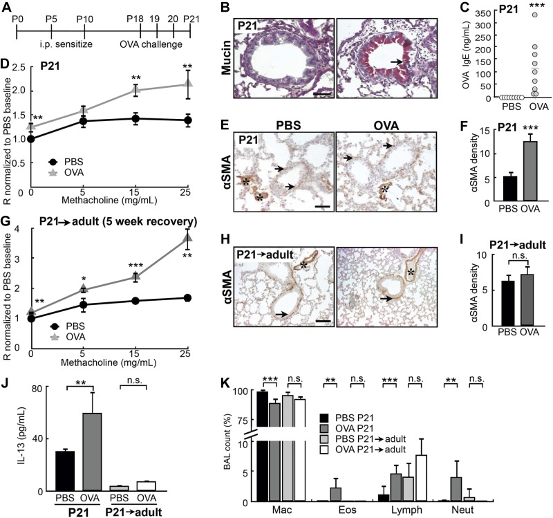
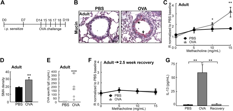
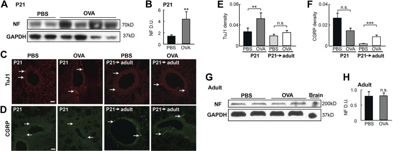
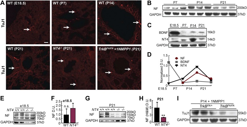
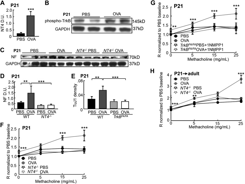
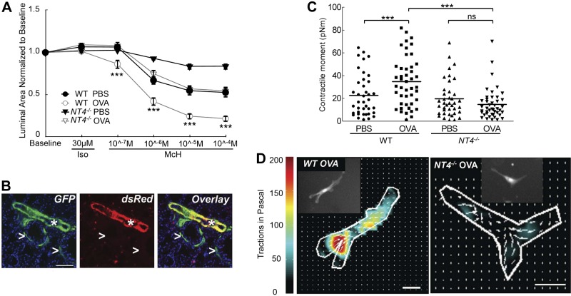
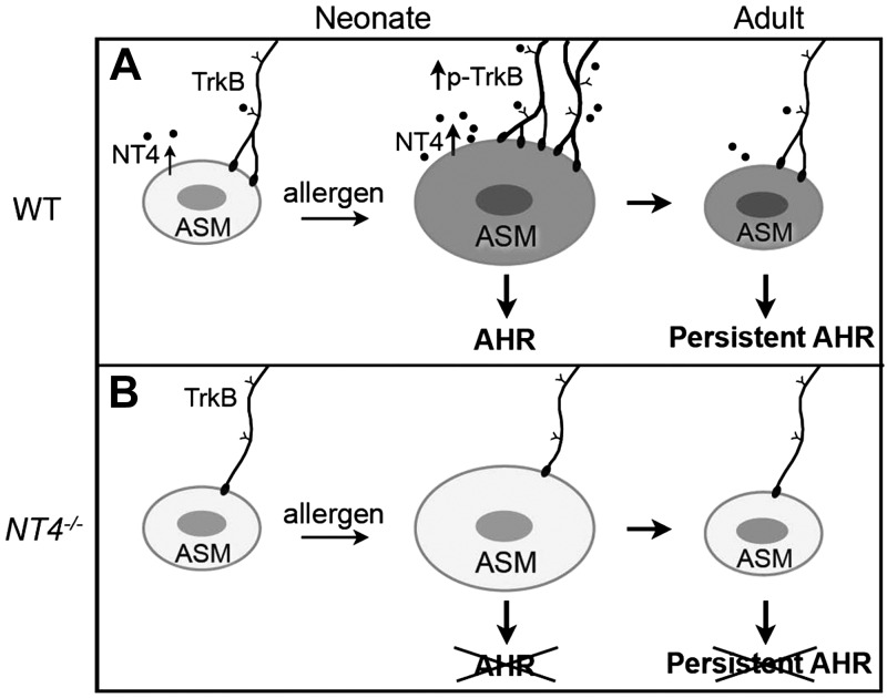
Children who are exposed to environmental respiratory insults often develop asthma that persists into adulthood. In this study, we used a neonatal mouse model of ovalbumin (OVA)-induced allergic airway inflammation to understand the long-term effects of early childhood insults on airway structure and function. We showed that OVA sensitization and challenge in early life led to a 2-fold increase in airway smooth muscle (ASM) innervation (P<0.05) and persistent airway hyperreactivity (AHR). In contrast, OVA exposure in adult life elicited short-term AHR without affecting innervation levels. We found that postnatal ASM innervation required neurotrophin (NT)-4 signaling through the TrkB receptor and that early-life OVA exposure significantly elevated NT4 levels and TrkB signaling by 5- and 2-fold, respectively, to increase innervation. Notably, blockade of NT4/TrkB signaling in OVA-exposed pups prevented both acute and persistent AHR without affecting baseline airway function or inflammation. Furthermore, biophysical assays using lung slices and isolated cells demonstrated that NT4 was necessary for hyperreactivity of ASM induced by early-life OVA exposure. Together, our findings show that the NT4/TrkB-dependent increase in innervation plays a critical role in the alteration of the ASM phenotype during postnatal growth, thereby linking early-life allergen exposure to persistent airway dysfunction.

Keywords: ASM; childhood asthma; contractility; innervation; lung slice; smooth muscle.


Figures

Figure 1.
Figure 1.
OVA exposure in early postnatal life led to persistent AHR. A) Experimental protocol for inducing allergic airway inflammation by OVA in a neonatal mouse model. Controls received saline (PBS) challenges. B) Mucin staining in the airways of PBS- and OVA-exposed pups at P21. Arrow indicates mucin+ cells. C) Serum levels of OVA-specific IgE in PBS- and OVA-exposed pups at P21, measured by ELISA. Each circle represents a sample. Red line indicates mean of the OVA-challenged group. D) Airway reactivity of control (n=14) and OVA-exposed (n=13) pups was assessed at P21 with the FlexiVent apparatus with increasing concentrations of methacholine. E) Representative images of αSMA immunostaining in the airways of PBS- and OVA-exposed pups at P21. Arrows indicate αSMA+ ASM; asterisks indicate αSMA blood vessels. F) Quantification of αSMA density in the airways of PBS- and OVA-exposed pups at P21. G) Control (n=9) and OVA-exposed (n=13) pups were allowed to grow for 5 wk into adulthood (P21→adult). Airway resistance was measured with the FlexiVent apparatus at 8 wk of age. H) Representative images of αSMA immunostaining in the airways of 8-wk-old adult mice (P21→adult) exposed to PBS and OVA as neonates. Arrows indicate αSMA+ ASM; asterisks indicate αSMA blood vessels. I) Quantification of αSMA density. J) Serum levels of IL-13 in PBS- and OVA-exposed pups at P21 and P21→adult mice, measured by ELISA. K) Differential cell count in BAL fluid of PBS- and OVA-exposed pups at P21 and P21→adult mice. Percentages of macrophages (Mac), eosinophils (Eos), lymphocytes (Lymph), and neutrophils (Neut) are shown. Scale bars = 50 μm. n.s., not significant. *P < 0.10; **P < 0.05; ***P < 0.01.
Figure 2.
Figure 2.
OVA exposure in adulthood did not lead to persistent AHR. A) Experimental protocol for inducing acute asthma in adult mice. Control mice received PBS challenges. B) Mucin staining in the airways of PBS- and OVA-exposed adult mice. Arrows indicate mucin+ cells. C) Airway reactivity of control (n=8) and OVA-challenged (n=5) adult mice was assessed at d 19 by measuring airway resistance with the FlexiVent apparatus. D) Quantification of αSMA density in the airways of PBS- and OVA-exposed adult mice. E) Serum levels of OVA-specific IgE in PBS- and OVA-exposed adult mice, measured by ELISA. Each circle represents a sample. Red line indicates mean of the OVA-challenged group. F) Airway reactivity of control and OVA-exposed mice was assessed 2.5 wk after the last challenge. G) Serum levels of IL-13 in PBS- and OVA-exposed adult mice at d 19 and after a 2.5 wk recovery, measured by ELISA. Scale bar = 50 μm. *P < 0.10; **P < 0.05; ***P < 0.01.
Figure 3.
Figure 3.
Early-life OVA exposure increased ASM innervation. A) Western blot analysis of NF in lung homogenates collected at P21 from controls and OVA-exposed pups. Each lane represents 1 mouse. B) Densitometry analysis of NF signal in the Western blot assay in (A). Data were normalized to the total amount of loaded protein. C) TuJ1 immunolabeling in airways of PBS- and OVA-exposed pups at P21 and in 8-wk-old mice exposed to PBS or OVA as neonates (P21→adult). Arrows indicate TuJ1-labeled axons. D) Immunostaining for CGRP in the airways of PBS- and OVA-exposed pups at P21 and in 8-wk-old adult mice exposed to PBS or OVA as neonates (P21adult). Arrows indicate CGRP-containing sensory afferents. E) Innervation density in control and OVA-exposed P21 pups and P21→adult mice. TuJ1 immunoreactivity was normalized to the size of each airway to calculate innervation density. Forty airways from 10 pups of each group were analyzed. F) Quantification of CGRP staining in the airways of P21 pups and P21→adult mice by normalizing CGRP immunoreactivity to the size of the airway. Forty airways from 10 pups of each group were analyzed. G) Western blot analysis of NF in lung homogenates of adult mice that were challenged with PBS or OVA. Lungs were harvested at d 19. H) NF levels were quantified by normalization to GAPDH levels. Data represent the average of 4 separate experiments with ≥3 mice/group for each experiment. Scale bars = 50 μm. n.s., not significant; **P < 0.05; ***P < 0.01.
Figure 4.
Figure 4.
NT4-activated TrkB signaling was necessary for ASM postnatal innervation. A) Representative images of TuJ1-labeled lung sections from WT mice before birth (E18.5) and at P7, P14, and P21. TuJ1 staining of lung sections from NT4−/− and 1NMPP1-treated TrkBFA/FA pups at P21 is also shown. Arrows indicate TuJ1-labeled axons in the airway. B) Western blot analysis of NF levels in lungs of pups at P7, P14, and P21. C) Western blot analysis of BDNF and NT4 levels in the lungs at E18.5, P7, P14, and P21. D) Densitometry analysis of NF, BDNF, and NT4 levels at E18.5 and during postnatal development, as shown in B and C. Signals were normalized to GAPDH levels. E) Western blot analysis of NF (high and medium molecular weight) levels in E18.5 whole-lung homogenates of WT and NT4−/− pups. F) NF signals, quantified by densitometry and normalized to GADPH levels. G) Western blot analysis of NF levels in P21 whole-lung homogenates of WT and NT4−/− pups. H) NF signals quantified and normalized. I) Western blot analysis of TuJ1 levels in P14 lungs of TrkBFA/FA pups and TrkBFA/+ littermates treated with 1NMPP1. J) TuJ1 signals, quantified and normalized to GADPH levels. Data represent results from 3 separate experiments with ≥3 mice/group for each experiment. Scale bar = 50 μm. n.s., not significant; **P < 0.05.
Figure 5.
Figure 5.
NT4-activated TrkB signaling was necessary for an early-life OVA-induced increase in lung innervation and AHR. A) NT4 levels in the lungs of controls and OVA-exposed pups at P21, analyzed by Western blot analysis and normalized to GAPDH levels. B) Western blot analysis of activated TrkB by phosphorylation in control and OVA-exposed lungs. C) Western blot analysis of NF at P21in WT and NT4−/− pups that were exposed to PBS and OVA. D) NF signals, quantified by densitometry and normalized to GAPDH levels. E) WT and TrkBFA/FA pups were exposed to PBS or OVA. TrkBFA/FA pups also received short-term 1NMPP1 treatment, to prevent OVA-induced overactivation of TrkB signaling. Innervation density in the lungs of each group was analyzed by TuJ1 immunolabeling. Graphs represent an average of 40 airways from 10 pups/group. F) Airway resistance of P21 WT and NT4−/− mice that were exposed to PBS or OVA (n=11/group). G) Airway resistance of WT and 1NMPP1-treated TrkBFA/FA pups that were exposed to PBS or OVA (WT-PBS, n=12; WT-Ova, n=9; TrkBFA/FA-PBS, n=9; and TrkBFA/FA-OVA, n=12). H) Airway resistance of adult WT and NT4−/− mice that had been exposed to PBS and OVA as pups (WT-PBS, n=9; NT4−/−-PBS; n=9; WT-OVA, n=13; and NT4−/−-OVA, n=16). Data represent the average of each group from 3 separate experiments. Data from the WT mice analyzed in F and G partially overlapped; results of WT mice at 8 wk of age in Fig. 1C were replotted in H, to allow a direct comparison between WT and NT4−/− mice. **P < 0.05; ***P < 0.01.
Figure 6.
Figure 6.
NT4 is required for OVA-induced increase in ASM contractile phenotypes. A) Measurement of methacholine-induced ASM contraction using lung slices. All lung slices were pretreated with isoproterenol to diminish differences in baseline contraction. A minimum of 26 airways from 3 mice were analyzed for each condition. There was a significant difference in lung slice contraction between OVA-exposed WT and NT4−/− mice. B) Differential GFP and dsRed expression in αSMA-GFP;NG2-dsRed pups at P21. Arrowheads indicate GFP+ ASM; asterisks indicate GFP+ and dsRed+ blood vessels. C) Contractile moment measurement of individual ASM cells based on their contraction map. ASM cells were isolated from αSMA-GFP;NG2-dsRed and NT4−/−SMA-GFP;NG2-dsRed pups after PBS and OVA exposure at P21. Each symbol represents the measurement of a single ASM cell; horizontal line represents the average within the group. D) Representative contraction maps of a single ASM cell isolated from lungs of OVA-exposed WT and NT4−/− pups. Insets: imaged GFP+ ASM cells. Scale bars = 50 μm. n.s., not significant. ***P < 0.01.
Figure 7.
Figure 7.
Model of how increased ASM innervation after early-life allergen exposure leads to persistent AHR. A) Postnatal lung innervation and early-life, allergen-induced AHR. NT4 is an ASM-derived neurotrophic factor for postnatal ASM innervation by TrkB-expressing extrinsic neurons. Early-life allergen exposure leads to ASM thickening and elevates NT4-activated TrkB signaling to increase ASM innervation. Increased innervation changes the ASM phenotype, ultimately resulting in persistent AHR without ASM thickening. B) Without NT4 signaling, ASM innervation in postnatal lung is reduced. In addition, although NT4 deficiency has no effect on ASM thickening after early-life allergen exposure, it blocks the increases in ASM innervation, which in turn prevents changes in ASM reactivity.

References

    1. Maddox L., Schwartz D. A. (2002) The pathophysiology of asthma. Annu. Rev. Med. 53, 477–498 - PubMed
    1. Stern D. A., Morgan W. J., Halonen M., Wright A. L., Martinez F. D. (2008) Wheezing and bronchial hyper-responsiveness in early childhood as predictors of newly diagnosed asthma in early adulthood: a longitudinal birth-cohort study. Lancet 372, 1058–1064 - PMC - PubMed
    1. Martinez F. D. (2009) The connection between early life wheezing and subsequent asthma: the viral march. Allergol. Immunopathol. (Madr.) 37, 249–251 - PubMed
    1. Bisgaard H., Bønnelykke K. (2010) Long-term studies of the natural history of asthma in childhood. J. Allergy Clin. Immunol. 126, 187–197 - PubMed
    1. Wu Z. X., Hunter D. D., Kish V. L., Benders K. M., Batchelor T. P., Dey R. D. (2009) Prenatal and early, but not late, postnatal exposure of mice to sidestream tobacco smoke increases airway hyperresponsiveness later in life. Environ. Health Perspect. 117, 1434–1440 - PMC - PubMed

Publication types