. 2014 Apr;32(4):886-901.
doi: 10.1002/stem.1599.

Wnt7b is an important intrinsic regulator of hair follicle stem cell homeostasis and hair follicle cycling

Affiliations

Wnt7b is an important intrinsic regulator of hair follicle stem cell homeostasis and hair follicle cycling

Eve Kandyba et al. Stem Cells. 2014 Apr.

Abstract

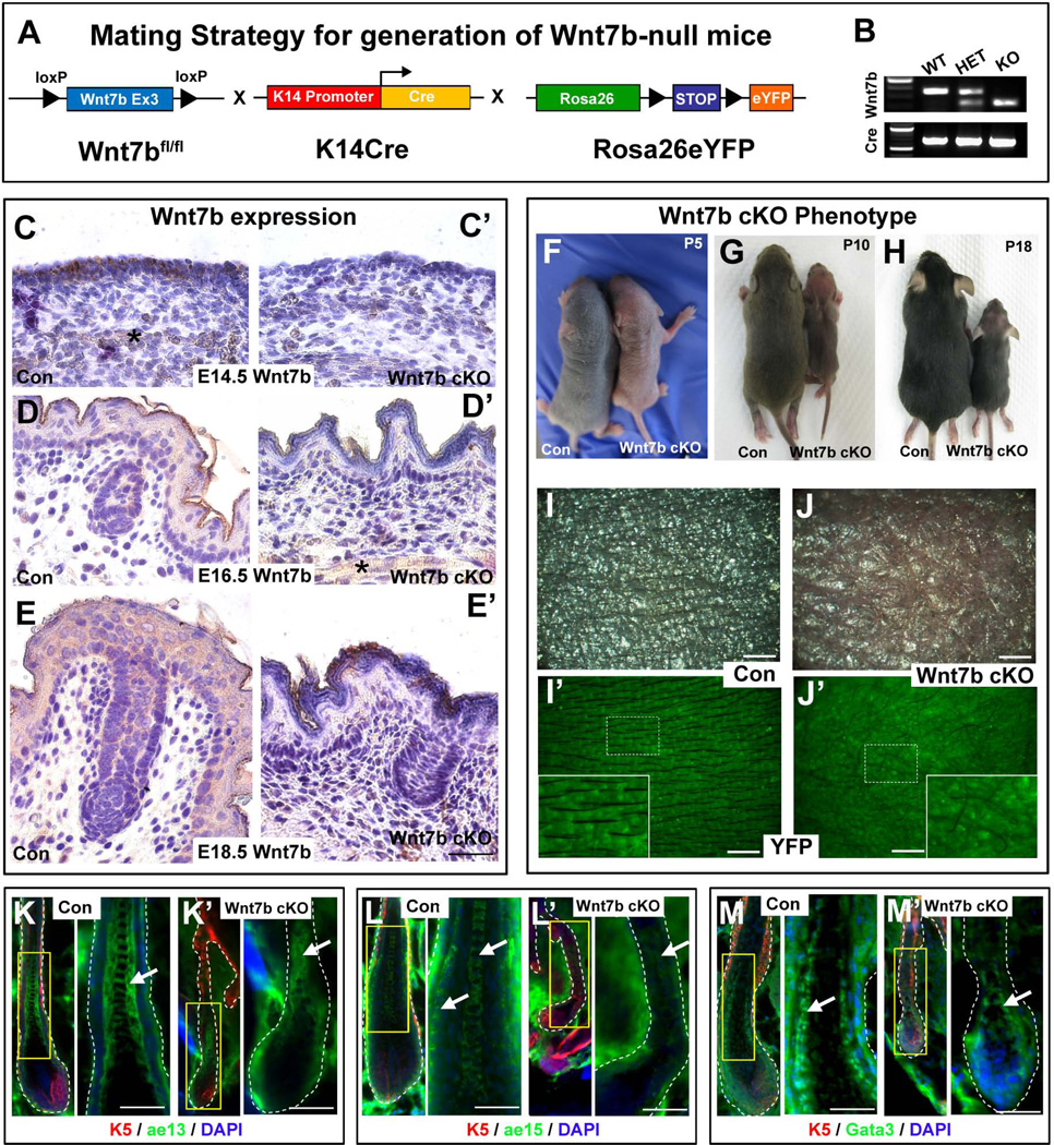
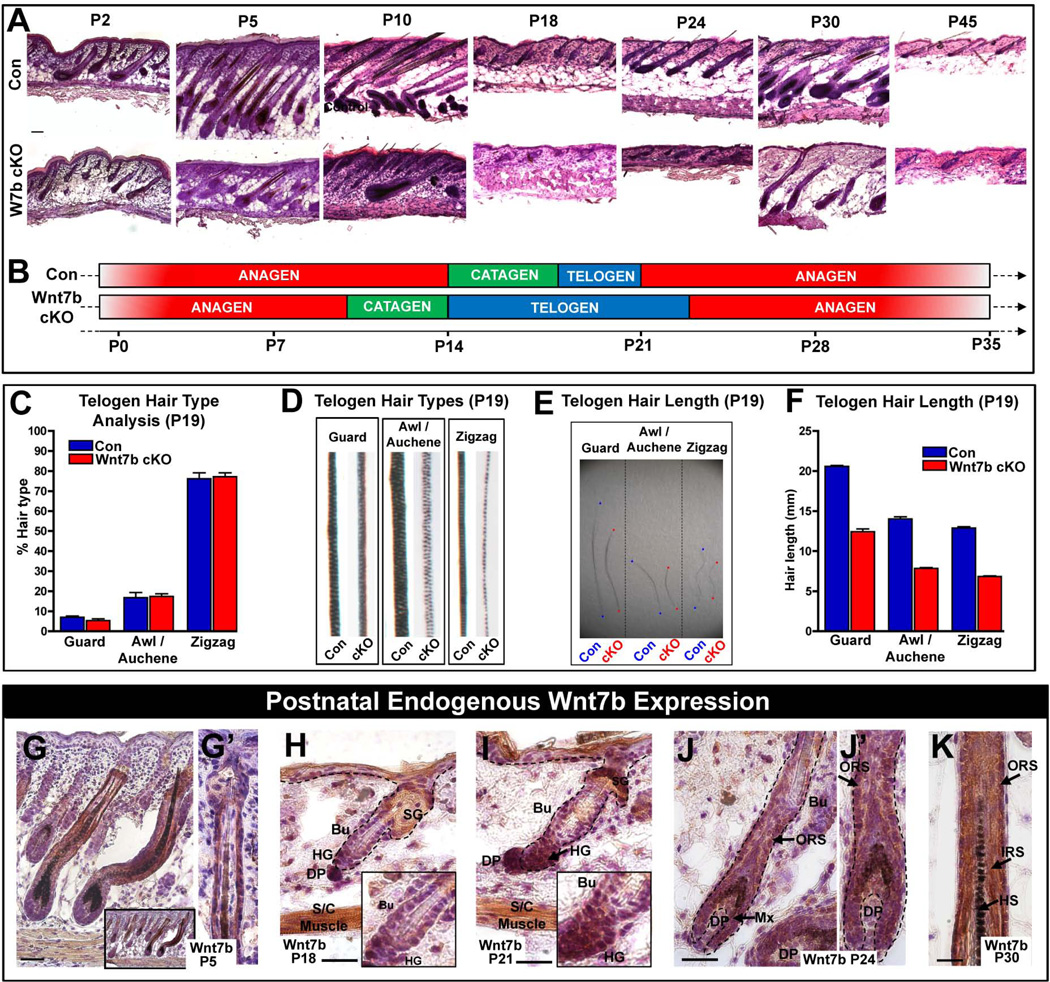
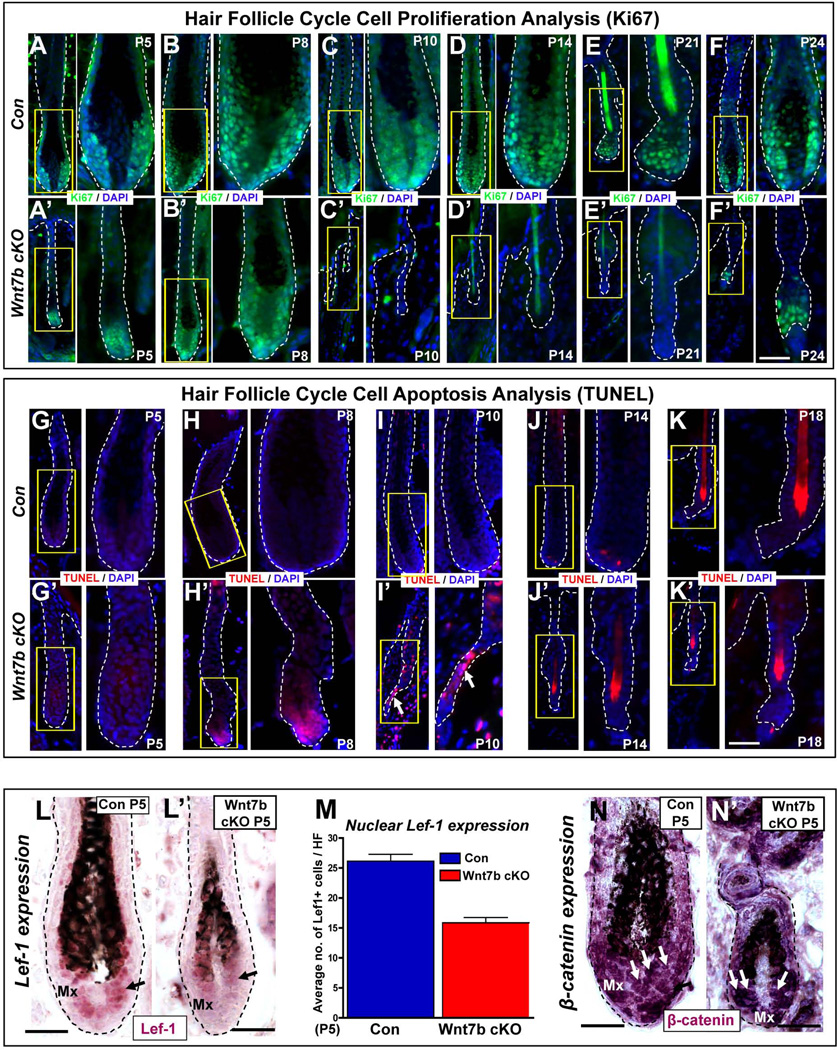
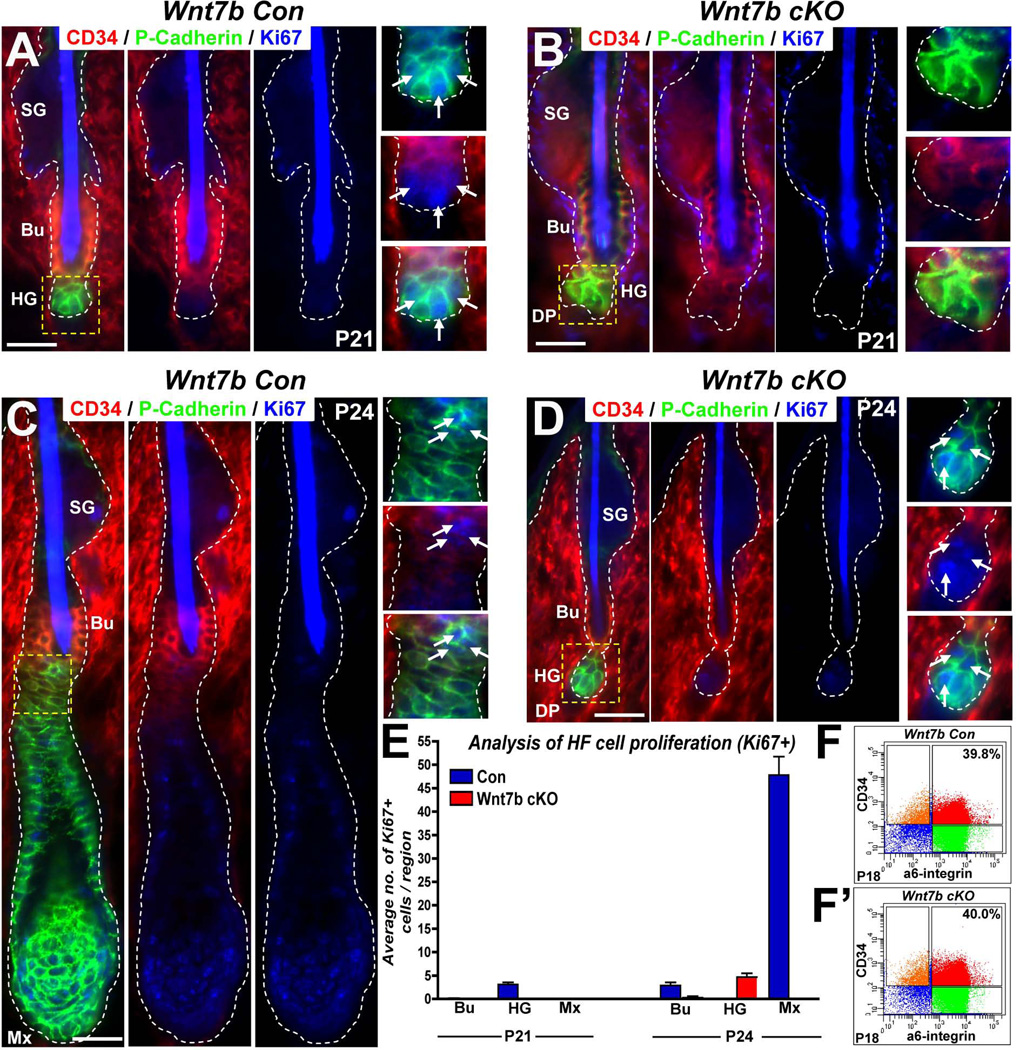
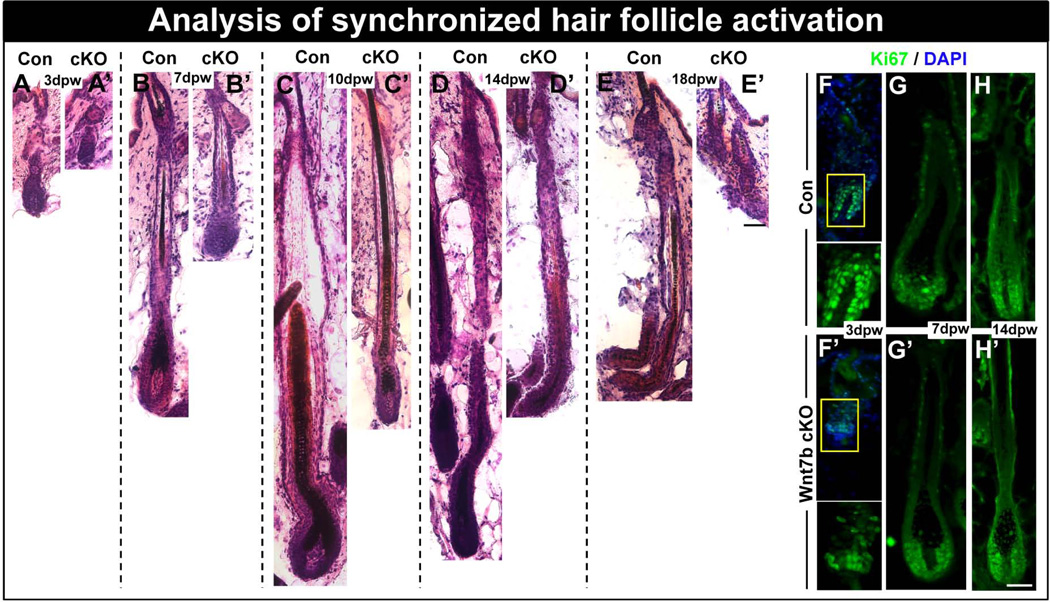
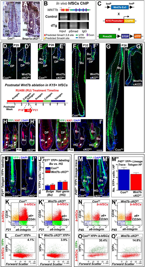
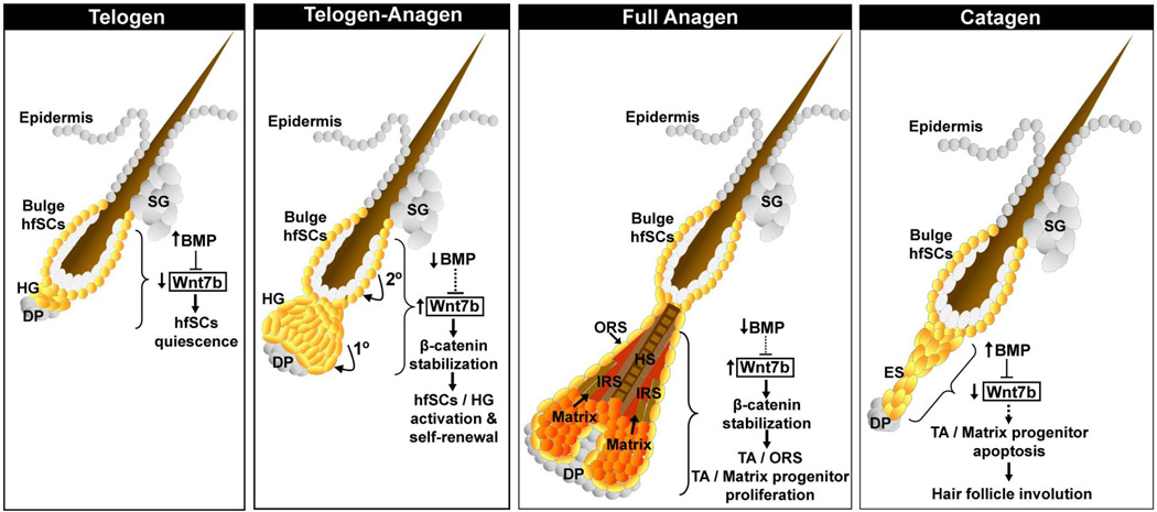
The hair follicle (HF) is an exceptional mini-organ to study the mechanisms which regulate HF morphogenesis, cycling, hair follicle stem cell (hfSCs) homeostasis, and progeny differentiation. During morphogenesis, Wnt signaling is well-characterized in the initiation of HF patterning but less is known about which particular Wnt ligands are required and whether individual Wnt ligands act in an indispensable or redundant manner during postnatal hfSCs anagen onset and HF cycle progression. Previously, we described the function of the bone morphogenetic protein (BMP) signaling target gene WNT7a in intrinsic regulation of hfSCs homeostasis in vivo. Here, we investigated the role of Wnt7b, which was also intrinsically upregulated in hfSCs during physiological and precocious anagen after BMP inhibition in vivo. We demonstrated Wnt7b to be a direct target of canonical BMP signaling in hfSCs and using Wnt7b conditional gene targeting during HF morphogenesis revealed disrupted HF cycling including a shorter anagen, premature catagen onset with overall shorter hair production, and diminished HF differentiation marker expression. Additionally, we observed that postnatal ablation of Wnt7b resulted in delayed HF activation, affecting both the hair germ and bulge hfSCs but still maintaining a two-step sequence of HF stimulation. Interestingly, Wnt7b cKO hfSCs participated in reformation of the new HF bulge, but with slower self-renewal. These findings demonstrate the importance of intrinsic Wnt7b expression in hfSCs regulation and normal HF cycling and surprisingly reveal a nonredundant role for Wnt7b in the control of HF anagen length and catagen entry which was not compensated by other Wnt ligands.

Keywords: Hair follicle stem cells, WNT signaling, Wnt7b.


Figures

Figure 1
Figure 1. Generation of Wnt7b-null (Wnt7b cKO) transgenic mice with aberrant hair coat production and diminished hair differentiation marker expression
(A) Schematic representation of the mating strategy used to generate Wnt7b-deficient transgenic mice. Wnt7bfl/fl transgenic mice were crossed with K14Cre mice and Rosa26-STOP-eYFP reporter mice to generate offspring with Cre-mediated deletion of Wnt7b in K14+ YFP-labeled epithelial tissue. (B) PCR genotyping was used to distinguish YFP+ control (Con) and YFP+ Wnt7b-deficient mice (Wnt7b cKO). (C, D, E) Immunohistochemistry (IHC) directed against endogenous Wnt7b protein was performed on Con and (C’, D’, E’) Wnt7b cKO dorsal back skin at E14.5, E16.5 and E18.5 to demonstrate efficient deletion of Wnt7b in Wnt7b cKO transgenic mouse skin. All IHC images were counterstained with hematoxylin (blue) to label nuceli. (F–H) Phenotype of Con and Wnt7b cKO transgenic mice at postnatal day (P) P5 (F), P10 (G) and P18 (H). (I, I’) Dorsal skin of P7 YFP+ Con mice displayed organized, visible hair shafts (HS) protruding from the skin surface. (J, J’) Dorsal skin of P7 YFP+ Wnt7b cKO transgenic mice displayed disorganized, aberrant and fewer hair shafts at the skin surface. Immunofluorescence directed against known hair follicle differentiation markers K5 (red, K–M’), ae13 (green, K, K’), ae15 (green; L, L’) and Gata3 (green, M, M’) in Con (K, L, M) and Wnt7b cKO transgenic (K’, L’, M’) hair follicles. DAPI (blue) counterstaining was used to label cell nuclei (K–M’). Asterisk labels subcutaneous muscle stained positive (brown) for Wnt7b. Scale bar = 50µm.
Figure 2
Figure 2. Wnt7b-deficient transgenic mice display aberrant hair follicle cycling
(A) Postnatal hair follicle (HF) cycle in Con and Wnt7b cKO transgenic mice (P5–45). (B) Schematic illustrating perturbed HF cycle in Wnt7b cKO mice compared with Con littermates. (C–F) Analysis of plucked, telogen (P19) hairs from Con and Wnt7b cKO mice. (C) Comparison of different HF types of Wnt7b cKO and Con mice. (D) Comparison of the hair shaft structure of Wnt7b cKO and Con mice. (E, F) Length of different hair types from Wnt7b cKO and Con HFs. (G, G’) Wnt7b expression in the ORS cells of P5 anagen HFs. (H, I and insets) Changes in Wnt7b expression during telogen to anagen transition between P18 and P21, strong cytoplasmic Wnt7b expression in the enlarged, activated HG and lower hfSCs (Bu) at anagen onset P21 compared to P18. (J, J’ magnification) At P24, during the first postnatal anagen, Wnt7b is expressed in the ORS, matrix progenitor cells and in the differentiating layers at P30 (K). All IHC images were counterstained with hematoxylin (blue) to label nuceli. Insets depict magnified regions within the figure. Abbreviations: P, postnatal day; DP, dermal papillae; SG, sebaceous gland; ORS, outer root sheath; HS, hair shaft; HFs; hair follicle; Bu, bulge; HG, hair germ; hfSCs, hair follicle stem cells; S/C. subcutaneous; IRS, inner root sheath. Scale bar = 50µm.
Figure 3
Figure 3. Wnt7b ablation results in a shorter hair cycle with premature catagen onset and delayed anagen entry
(A–F’) Immunofluorescence targeting the cell proliferation marker, Ki67 (green) during the postnatal hair cycle (from P5–P24) in Con (A–F) and Wnt7b cKO (A’–F’) mice. TUNEL staining (red) was used to detect cell apoptosis and indicate initiation of the degenerative hair follicle phase, catagen, in Con (G–K) and Wnt7b cKO (G’–K’) hair follicles. DAPI (blue) counterstaining was used to label cell nuclei. Arrow marks the TUNEL positive, degenerating epithelial strand. Immunohistochemistry (IHC) detecting nuclear Lef-1 (purple) in Con (L) and Wnt7b cKO (L’) at P5 with quantification of the number of nuclear Lef-1 positive cells per HF (M). (N–N’) IHC targeting β-catenin expression in P5 Con (N) and Wnt7b cKO (N’) HFs. Scale bar = 50µm.
Figure 4
Figure 4. Wnt7b-deficiency does not perturb the sequential “two-step” mechanism of HF activation during telogen-anagen transision
(A–D) Triple immunofluorescence (IF) targeting cell proliferation (Ki67, blue) in the bulge (Bu) hfSCs using CD34 (red) marker expression and the hair germ (HG) using P-cadherin (green) staining in Con (A, C) and Wnt7b cKO (B, D) HFs at P21 (A–B) and P24 (C–D). (E) Quantification of nuclear Ki67 IF in specific HF compartments (Bu, bulge hfSCs, HG, hair germ; Mx, matrix cells) at P21 and P24. (F–F’) FACS analysis of P18 Con (F) and Wnt7b cKO (F’) HFs employing CD34 and α6-integrin marker expression. α6-integrinHigh/CD34High (red) denotes comparable total basal hfSCs (b-hfSCs) numbers in the bulge of Con (E, 39.8%) and Wnt7b cKO (E’, 40%) HFs.
Figure 5
Figure 5. Wnt7b-deficient hair follicles display delayed anagen entry after hair cycle synchronization by depilation
(A–E’) Con and Wnt7b cKO dorsal hair was waxed to synchronize hair follicle activation (promoting anagen entry) and hair re-growth monitored and stained by H&E. (A) After 3 days postwaxing (pw) Con HFs revealed HF downgrowth in early anagen with HFs extending into the subcutaneous fat layer, however, at the same time point (A’), Wnt7b cKO HF regrowth appeared delayed and remained more superficial to the epidermis. (B) At 7 days pw, Con anagen HFs extended deep into the subcutaneous fat tissue (B’) whereas, Wnt7b cKO still remained delayed by comparison. (C) After 10 days pw, Con full anagen HFs produced thick, pigmented hair shafts, (C’) however, Wnt7b cKO HFs appeared considerably thinner with delayed downgrowth into the dermis. (D) 14 days pw, Con HFs remained in anagen (D'), however, Wnt7b cKO HFs were in early catagen. (E) At 18 days pw, the lower portion of Con HFs were beginning to degenerate in catagen, whereas, Wnt7b cKO HFs had already transitioned to telogen (E’). (F–H’) Immunofluorescence detecting Ki67 (green) in regenerating Con (F–H) and Wnt7b cKO (F’–H’) HFs at 3dpw (F, F’), 7dpw (G, G’) and 14dpw (H, H’). DAPI (blue) counterstaining was used to label cell nuclei in immunofluorescence images. Scale bar = 50µm.
Figure 6
Figure 6. Delayed hair follicle stem cell (hfSCs) activation following Wnt7b ablation
(A’) Up-regulation of WNT7b in telogen bulge (Bu) hfSCs and hair germ (HG) after BMP pathway inactivation in BMPR1A cKORU at P59 compared to ConRU HFs (A) where WNT7b staining was absent. (B) In vivo hfSCs ChIP PCR reveals selective precipitation of DNA fragments that possess canonical Smad binding elements of Wnt7b promoter, flanking primers (arrows) in schematic (C) Schematic of transgenic mice generation with targeted Wnt7b deletion in the bulge: Con and Wnt7bfl/fl transgenic mice (both K15CrePR+; YFP+) were treated with RU486 from P18–P21 (schematic inset) to YFP label K15+ control (ConRU) and ablate Wnt7b in Wnt7b cKORU hfSCs. (D–G’) The following postnatal hair cycle was monitored. (D, D’) After 4 days (P25), both ConRU and Wnt7b cKORU HFs remained in telogen due to RU treatment . (E) By P28, YFP+ ConRU HFs displayed telogen-anagen transition with enlarged, activated HG, however, YFP+ Wnt7b cKORU HFs remained in telogen (E’). At P32, YFP+ ConRU HFs were in full anagen with efficient YFP+ labeling throughout all HF layers (F), however, YFP+ Wnt7b cKORU displayed enlarged HGs indicating initial HF activation and early telogen-anagen transition (F’). By P36, ConRU YFP+ HFs were in full anagen with HFs extending deep into the dermis (G), whereas, Wnt7b cKORU YFP+ HFs were in early anagen (G’). (H–I’) Immunofluorescence for the cell proliferation marker, Ki67 in YFP+ ConRU and Wnt7b cKORU HFs. (H) At P28, ConRU YFP+ HFs display strong nuclear Ki67 staining in the HG and lower Bu cells indicating HF activation, (H’) however, Wnt7b cKORU HFs lacked Ki67 staining. (I) At P32, nuclear Ki67 staining was detected in the matrix cells of YFP+ ConRU HFs. (I’) Wnt7b cKORU YFP+ HFs displayed nuclear Ki67 staining in the HG indicating early HF activation. DAPI (blue) counterstaining was used to label all nuclei. Representative images of P21 ConDil (J) and Wnt7b cKODil (J’) YFP+ labeled HFs and quantification of the number of YFP+ cells per Bu and HG compartment (J”). (K–K’) FACS analysis employing α6-integrin and CD34 staining with α6-integrinHigh/CD34High basal hfSCs population (b-hfSCs, red) in ConDil and Wnt7b cKODil at P21 HFs. FACS analysis of b-hfSCs (α6High/CD34High) gated population for YFP positive cells in Con (L) and Wnt7b cKO (L’) at P21 HFs. Representative images of P45 ConDil (M) and Wnt7b cKODil (M’) YFP+ labeled HFs and quantification of the number of YFP+ cells per HF (M”). (N–N’) FACS analysis of α6-integrin and CD34 positive b-hfSCs population (b-hfSCs, red; α6-integrinHigh/CD34High) in ConDil and Wnt7b cKODil at P45 HFs. FACS analysis of b-hfSCs (α6High/CD34High) gated population for YFP positive cells in ConDil (O) and Wnt7b cKODil (O’) at P45 HFs. Scale bar = 50µm.
Figure 7
Figure 7. Model of the intrinsic role of Wnt7b and cross talk with BMP signaling in postnatal hair follicle stem cells (hfSCs) regulation and postnatal hair follicle cycling
Schematic model summarizing the role of Wnt7b in hfSCs during telogen and the telogen-anagen transition. During telogen-anagen transition, Wnt7b promotes a two-step sequence of HF stimulation, with initial activation in the HG (1°) and subsequently in the bulge hfSCs (2°). Proposed role for Wnt7b in matrix progenitor cells during the growth (anagen) and degenerative (catagen) phase of the postnatal hair follicle cycle. Loss of Wnt7b expression may indirectly be sufficient to promote HF involution by diminishing the collective WNT signaling during catagen (dotted arrow).

References

    1. Hardy MH. The secret life of the hair follicle. Trends Genet. 1992;8:55–61. - PubMed
    1. Millar SE. Molecular mechanisms regulating hair follicle development. J Invest Dermatol. 2002;118:216–225. - PubMed
    1. Sengel P, Mauger A. Peridermal cell patterning in the feather-forming skin of the chick embryo. Dev Biol. 1976;51:166–171. - PubMed
    1. Muller-Rover S, Peters EJ, Botchkarev VA, et al. Distinct patterns of NCAM expression are associated with defined stages of murine hair follicle morphogenesis and regression. J Histochem Cytochem. 1998;46:1401–1410. - PubMed
    1. Blanpain C, Lowry WE, Geoghegan A, et al. Self-renewal, multipotency, and the existence of two cell populations within an epithelial stem cell niche. Cell. 2004;118:635–648. - PubMed

Publication types

MeSH terms