

doi: 10.1155/2013/924902.
Epub 2013 Oct 9.
Vulvar fibroadenoma with lactational changes in ectopic breast tissue
Affiliations
- PMID: 24222874
- PMCID: PMC3810074
- DOI: 10.1155/2013/924902
Item in Clipboard
Vulvar fibroadenoma with lactational changes in ectopic breast tissue
Case Rep Obstet Gynecol.
2013.
Abstract
Ectopic breast tissue represents any type of breast tissue found outside its normal location in the pectoral region. The second most common location for ectopic breast tissue after axilla is the vulvar region. We present a case of a healthy 20-year-old female, G1P1, who presented to the Emergency Department with a sudden increase in size of a painful mass located in her vulva, which started 4 days after a spontaneous vaginal delivery and 3 days after initiation of breast-feeding of her newborn. She reported a stable, smaller, painless mass in the same location for almost 2 years prior to this episode. After surgical excision, a fibroadenoma with lactation changes within ectopic breast tissue was confirmed.
Figures
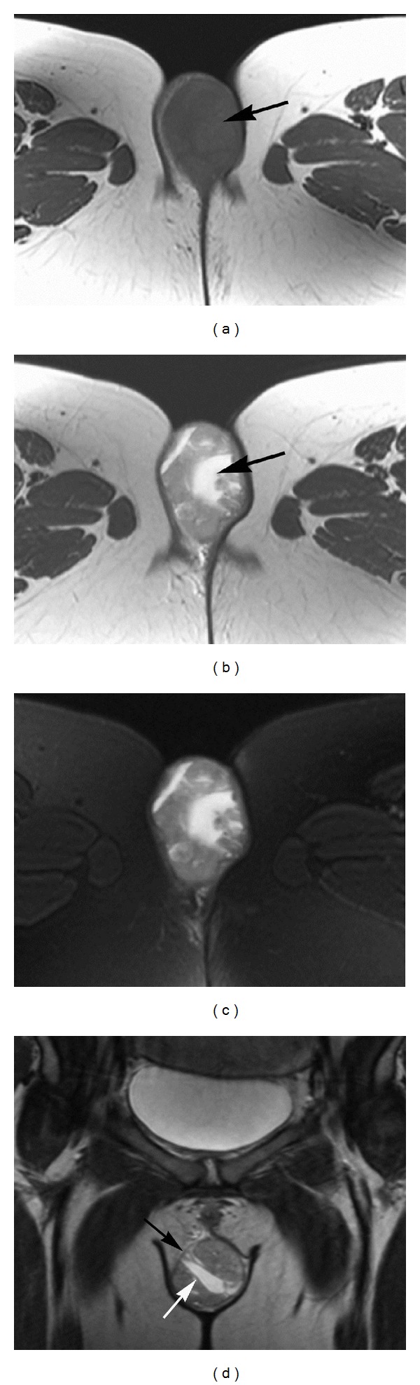
Magnetic resonance images of the pelvis. (a) Axial T1-weighted spin-echo image (TR = 700 ms/TE = 10 ms). A well-defined, ovoid mass in the subcutaneous tissue of the inferior right labia majora is seen (arrow), with smooth contours and no evidence of infiltration of adjacent structures. The mass has homogenous signal which is isointense to skeletal muscle. (b) Axial T2-weighted fast spin-echo image (TR = 4450 ms/TE = 89 ms). The mass has heterogeneous high signal intensity, with areas of very high signal intensity suggesting the presence of fluid (arrow) (c) Axial T2-weighted fast spin-echo image with frequency selective fat saturation (TR = 5266 ms/TE = 89 ms). No inflammatory changes are seen in the adjacent fat. (d) Coronal T2-weighted fast spin-echo image (TR = 5216 ms/TE = 83 ms). A well-defined thin capsule around the mass (black arrow) and thin internal septa (white arrow) are demonstrated.

Histology slides of the lesion show (a) lobular architecture and associated ischemic necrosis (arrow). (b) Epithelial and mesenchymal proliferation with apocrine change (arrow). (c) Focal squamous metaplasia (arrow). Images were acquired using H&E staining with 40x (a) and 100x ((b), (c)) magnification.
References
-
- Lee E-S, Kim I. Multiple vulvar lactating adenomas. Obstetrics and Gynecology. 2011;118(2):478–480. - PubMed
-
- Tüzün YO, Engin B, Wolf R. Distribution and arrangement of multiple lesions in the anogenital region. Clinics in Dermatology. 2011;29(2):162–172. - PubMed
-
- Di Gilio AR, Cormio G, Resta L, et al. Rapid growth of myxoid leiomyosarcoma of the vulva during pregnancy: a case report. International Journal of Gynecological Cancer. 2004;14(1):172–175. - PubMed
-
- Baker TP, Lenert JT, Parker J, et al. Lactating adenoma: a diagnosis of exclusion. Breast Journal. 2001;7(5):354–357. - PubMed
-
- Irvin WP, Cathro HP, Grosh WW, Rice LW, Andersen WA. Primary breast carcinoma of the vulva: a case report and literature review. Gynecologic Oncology. 1999;73(1):155–159. - PubMed
LinkOut - more resources
Full Text Sources
Other Literature Sources
