

Spontaneous intramural duodenal hematoma in type 2B von Willebrand disease
- PMID: 24222967
- PMCID: PMC3819559
- DOI: 10.3748/wjg.v19.i41.7205
Spontaneous intramural duodenal hematoma in type 2B von Willebrand disease
Abstract
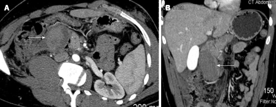
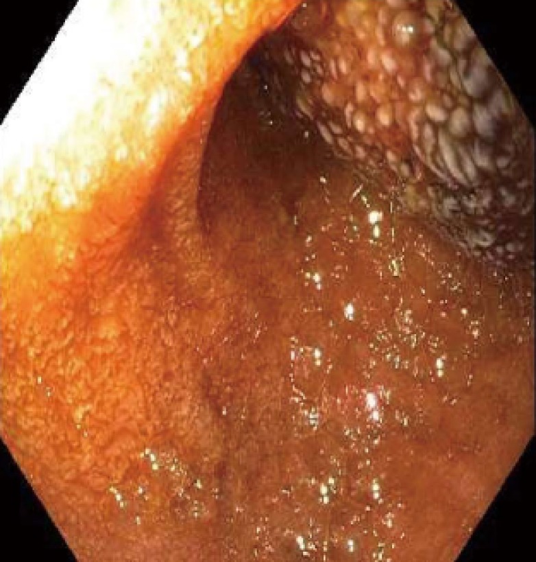
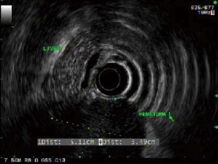
Intramural duodenal hematoma is a rare cause of a proximal gastrointestinal tract obstruction. Presentation of intramural duodenal hematoma most often occurs following blunt abdominal trauma in children, but spontaneous non-traumatic cases have been linked to anticoagulant therapy, pancreatitis, malignancy, vasculitis and endoscopy. We report an unusual case of spontaneous intramural duodenal hematoma presenting as an intestinal obstruction associated with acute pancreatitis in a patient with established von Willebrand disease, type 2B. The patient presented with abrupt onset of abdominal pain, nausea, and vomiting. Computed tomography imaging identified an intramural duodenal mass consistent with blood measuring 4.7 cm × 8.7 cm in the second portion of the duodenum abutting on the head of the pancreas. Serum lipase was 3828 units/L. Patient was managed conservatively with bowel rest, continuous nasogastric decompression, total parenteral nutrition, recombinant factor VIII (humateP) and transfusion. Symptoms resolved over the course of the hospitalization. This case highlights an important complication of an inherited coagulopathy.
Keywords: Duodenal hematoma; von Willebrand disease.
Figures
References
-
- Hoenisch K, Prommegger R, Schwaighofer H, Freund M, Schocke M, Vogel W, Kaser A. Intramural duodenal hematoma after upper gastrointestinal endoscopy. Wien Med Wochenschr. 2011;161:441–444. - PubMed
-
- Weil BR, Howard TJ, Zyromski NJ. Spontaneous duodenal hematoma: a rare cause of upper gastrointestinal tract obstruction. Arch Surg. 2008;143:794–796. - PubMed
-
- Hou SW, Chen CC, Chen KC, Ko SY, Wong CS, Chong CF. Sonographic diagnosis of spontaneous intramural small bowel hematoma in a case of warfarin overdose. J Clin Ultrasound. 2008;36:374–376. - PubMed
-
- McLachlan J. Fatal false aneurismal tumor occupying nearly the whole of the duodenum. Lancet. 1838;2:203–205.
Publication types
MeSH terms
Substances
LinkOut - more resources
Full Text Sources
Other Literature Sources
Medical
Miscellaneous
