. 2013 Oct 25;8(10):e78542.
doi: 10.1371/journal.pone.0078542. eCollection 2013.

New strategies to prolong the in vivo life span of iron-based contrast agents for MRI

Affiliations

New strategies to prolong the in vivo life span of iron-based contrast agents for MRI

Antonella Antonelli et al. PLoS One. .

Abstract

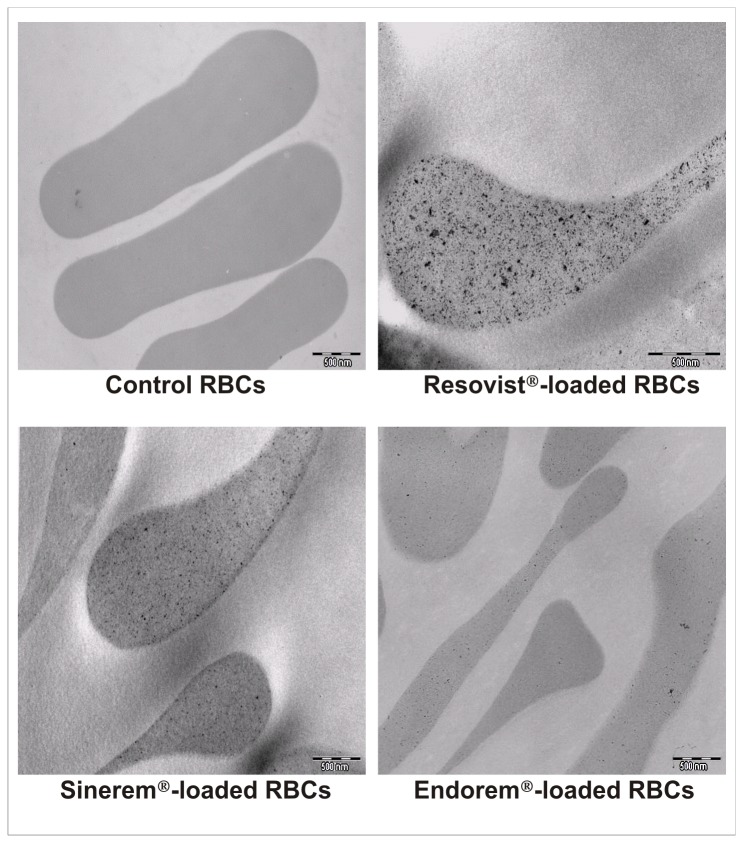
Superparamagnetic iron oxide (SPIO) and ultra small superparamagnetic iron oxide (USPIO) nanoparticles have been developed as magnetic resonance imaging (MRI) contrast agents. Iron oxide nanoparticles, that become superparamagnetic if the core particle diameter is ~ 30 nm or less, present R1 and R2 relaxivities which are much higher than those of conventional paramagnetic gadolinium chelates. Generally, these magnetic particles are coated with biocompatible polymers that prevent the agglomeration of the colloidal suspension and improve their blood distribution profile. In spite of their potential as MRI blood contrast agents, the biomedical application of iron oxide nanoparticles is still limited because of their intravascular half-life of only few hours; such nanoparticles are rapidly cleared from the bloodstream by macrophages of the reticulo-endothelial system (RES). To increase the life span of these MRI contrast agents in the bloodstream we proposed the encapsulation of SPIO nanoparticles in red blood cells (RBCs) through the transient opening of cell membrane pores. We have recently reported results obtained by applying our loading procedure to several SPIO nanoparticles with different chemical physical characteristics such as size and coating agent. In the current investigation we showed that the life span of iron-based contrast agents in the mice bloodstream was prolonged to 12 days after the intravenous injection of murine SPIO-loaded RBCs. Furthermore, we developed an animal model that implicates the pretreatment of animals with clodronate to induce a transient suppression of tissue macrophages, followed by the injection of human SPIO-loaded RBCs which make it possible to encapsulate nanoparticle concentrations (5.3-16.7 mM Fe) higher than murine SPIO-loaded RBCs (1.4-3.55 mM Fe). The data showed that, when human RBCs are used as more capable SPIO nanoparticle containers combined with a depletion of tissue macrophages, Fe concentration in animal blood is 2-3 times higher than iron concentration obtained by the use of murine SPIO-loaded RBCs.


Conflict of interest statement

Competing Interests: M. Magnani declares stock ownership in EryDel SpA. Guerbet provided the Sinerem® contrast for this study. This does not alter the authors' adherence to all the PLOS ONE policies on sharing data and materials, as detailed online in the guide for authors.

Figures

Figure 1
Figure 1. Relaxivity constants r1 or r2 of SPIO nanoparticles added to RBCs.
r1 (A) and r2 (B) relaxivity constants of SPIO nanoparticles added to human RBCs. r1 (C) and r2 (D) relaxivity constants of SPIO nanoparticles added to murine RBCs. Exact r1 and r2 values are reported in parentheses next to the name of the material encapsulated in the RBCs.
Figure 2
Figure 2. Transmission Electron Microscopy (TEM) analysis of human SPIO-loaded RBCs.
TEM images show the presence of magnetic nanoparticles in RBCs loaded with Resovist®, Sinerem® or Endorem® contrast agents compared to unloaded control cells.
Figure 3
Figure 3. In vivo pharmacokinetic NMR results after intravenous injection in mice of murine Resovist®-loaded RBCs.
(A) T1 values of blood samples from treated and control mice. (B) The percentage of Fe μmoles in the blood circulatory system compared to the Fe dose injected or by Resovist®-loaded RBCs or as free Resovist®. Values are expressed as means of three experiments and the error bars show the standard deviations. Asterisks indicate statistical significance; (ns) not significant; (**) p<0.01 and (***) p<0.001.
Figure 4
Figure 4. Histological sections of ICR (CD-1®) mice liver and spleen tissues stained by Perl’s method.
Iron presence in liver and spleen sections was detected after the i.v. injection of human unloaded- or SPIO-loaded RBCs in mice treated or not treated with clodronate. Group I: control mice. Group II: mice receiving unloaded RBCs. Group III: mice pretreated with clodronate receiving unloaded RBCs. Group IV: mice receiving Sinerem®-loaded RBCs. Group V: mice pretreated with clodronate and receiving Sinerem®-loaded RBCs. Magnification 20X.
Figure 5
Figure 5. AAS analyses of Fe content in kidney, liver and spleen tissues of ICR
(CD-1®) mice. The iron content in the organs of mice treated or not treated with clodronate before human unloaded- or Sinerem®-loaded RBCs intravenously administered was analysed by atomic absorption spectrometry. Group I: control mice. Group II: mice receiving unloaded RBCs. Group III: mice pretreated with clodronate receiving unloaded RBCs. Group IV: mice receiving Sinerem®-loaded RBCs. Group V: mice pretreated with clodronate receiving Sinerem®-loaded RBCs. Values are expressed as means of three experiments and the error bars show the standard deviations. Asterisks indicate statistical significance versus control group (Group I); (ns) not significant; (*) p ≤ 0.05, (**) p<0.01 and (***) p<0.001.
Figure 6
Figure 6. Histological sections of ICR (CD-1®) mice liver and spleen tissues marked with F4/80 antibody.
Livers and spleens were removed 24 hours post human RBC injection and the F4/80 monoclonal antibody was used as a marker for mouse macrophages. Brown patches represent stained macrophages. Group I: control mice. Group II: mice receiving unloaded RBCs. Group III: mice pretreated with clodronate receiving unloaded RBCs. Group IV: mice receiving Sinerem®-loaded RBCs. Group V: mice pretreated with clodronate receiving Sinerem®-loaded RBCs. Magnification 40X.
Figure 7
Figure 7. Macrophage depletion in liver and spleen tissues of ICR (CD-1®) mice.
The number of liver (A) and spleen (B) macrophages was evaluated by counting ten fields/slides 24 hours after human unloaded- or SPIO-loaded RBC administration. The results are expressed in percentage compared to untreated control mice. Values are expressed as means of three experiments and the error bars show the standard deviations. Asterisks indicate statistical significance versus control group (Group I); (ns) not significant; (*) p ≤ 0.05, (**) p<0.01 and (***) p<0.001.
Figure 8
Figure 8. Pharmacokinetics of human Sinerem®-loaded RBCs in Sprague Dawley® rats.
(A) Percentage of Fe µmoles in the bloodstream of rats calculated in comparison with the injected dose after T1 NMR measurements. (B) Cytometric determinations of human RBC percentages present in the rat bloodstream after human unloaded- and Sinerem®-loaded RBC intravenous injection. Group II: rats receiving human unloaded RBCs. Group III: rats receiving human Sinerem®-loaded RBCs. Group IV: clodronate pretreated rats receiving Sinerem®-loaded RBCs. Values are expressed as means of three experiments and the error bars show the standard deviations. Asterisks indicate statistical significance; (ns) not significant; (*) p≤ 0.05; (**) p<0.01 and (***) p<0.001.
Figure 9
Figure 9. Cytometric detection of human SPIO-loaded RBCs in rat blood circulation.
Contour plot FSC vs Anti-A FITC fluorescence of red blood cells from rats receiving: unloaded RBCs (Group II, left column), Sinerem®-loaded RBCs (Group III, middle column), and Sinerem®-loaded RBCs after clodronate pretreatment (Group IV, right column). Images are related to cytometry analyses of blood samples taken at 24h (A, B, C), 48h (D, E, F) and 5 days (G, H, I). The position of bars was established taking into account cellular self-fluorescence on fluorescence profile (FL1). Percentages of human RBCs positive for anti-A labelling are specified in the figures.

References

    1. Rümenapp C, Gleich B, Haase A (2012) Magnetic nanoparticles in magnetic resonance imaging and diagnostics. Pharm Res 29: 1165-1179. doi:10.1007/s11095-012-0711-y. PubMed: 22392330. - DOI - PubMed
    1. Wang YX (2011) Superparamagnetic iron oxide based MRI contrast agents: Current status of clinical application. Quant Imaging Med Surg 1: 35-40. PubMed: 23256052. - PMC - PubMed
    1. Neuberger T, Schopf B, Hofmann H, Hofmann M, Von Rechenberg B (2005) Superparamagnetic nanoparticles for biomedical applications: Possibilities and limitations of a new drug delivery system. J Magn Magn Mater 293: 483-496. doi:10.1016/j.jmmm.2005.01.064. - DOI
    1. Weissleder R, Elizondo G, Wittenberg J, Rabito CA, Bengele HH et al. (1990) Ultrasmall superparamagnetic iron oxide: characterization of a new class of contrast agents for MR imaging. Radiology 175(2): 489-493. PubMed: 2326474. - PubMed
    1. Bulte JWM, Kraitchman DL (2004) Iron oxide MR contrast agents for molecular and cellular imaging. NMR Biomed 17(7): 484–499. doi:10.1002/nbm.924. PubMed: 15526347. - DOI - PubMed

Publication types