. 2013 Nov 6;8(11):e79958.
doi: 10.1371/journal.pone.0079958. eCollection 2013.

Growth and airborne transmission of cell-sorted life cycle stages of Pneumocystis carinii

Affiliations

Growth and airborne transmission of cell-sorted life cycle stages of Pneumocystis carinii

Anna Martinez et al. PLoS One. .

Abstract

Pneumocystis organisms are airborne opportunistic pathogens that cannot be continuously grown in culture. Consequently, the follow-up of Pneumocystis stage-to-stage differentiation, the sequence of their multiplication processes as well as formal identification of the transmitted form have remained elusive. The successful high-speed cell sorting of trophic and cystic forms is paving the way for the elucidation of the complex Pneumocystis life cycle. The growth of each sorted Pneumocystis stage population was followed up independently both in nude rats and in vitro. In addition, by setting up a novel nude rat model, we attempted to delineate which cystic and/or trophic forms can be naturally aerially transmitted from host to host. The results showed that in axenic culture, cystic forms can differentiate into trophic forms, whereas trophic forms are unable to evolve into cystic forms. In contrast, nude rats inoculated with pure trophic forms are able to produce cystic forms and vice versa. Transmission experiments indicated that 12 h of contact between seeder and recipient nude rats was sufficient for cystic forms to be aerially transmitted. In conclusion, trophic- to cystic-form transition is a key step in the proliferation of Pneumocystis microfungi because the cystic forms (but not the trophic forms) can be transmitted by aerial route from host to host.


Conflict of interest statement

Competing Interests: The authors have declared that no competing interests exist.

Figures

Figure 1
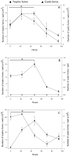
Figure 1. Growth of sorted P. carinii organisms in axenic culture.
Several P. carinii populations were cultured in DMEM with 10% FBS, at 37°C, in an atmosphere containing 5% CO2. Growth of (A) sorted P. carinii total population devoid of host cell debris, (B) pure P. carinii trophic forms, and (C) pure P. carinii cystic forms is followed during 4 days. Trophic forms (circles, left Y-axis) or cystic forms (triangles, right Y-axis) were microscopically quantified after RAL-555 panoptic staining [26-28]. For each population of P. carinii organisms studied, means of three replicates are represented per time point. Error bars represent standard deviations. The star (*) means P-value ≤ 0.05. The detection limit is 10 P. carinii organisms per culture well.
Figure 2
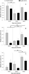
Figure 2. Infection of nude rats with sorted P. carinii organisms.
Nude rats were endotracheally infected with three P. carinii sorted populations: (A) sorted P. carinii total population devoid of host cell debris, (B) pure P. carinii trophic forms, and (C) pure P. carinii cystic forms. Rats were euthanized at 0.5 (12 h), 2.5 days, and 8.5 days postinoculation and then parasites were extracted from lungs [29]. After staining with RAL-555, trophic forms (in grey) or cystic forms (in black) were microscopically quantified [26-28]. At each time point, means of either trophic- or cystic-form burdens developing in the lungs of three animals are plotted on the same Y-axis. Error bars represent standard deviations. The star (*) means P-value ≤ 0.05.
Figure 3
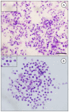
Figure 3. Purity of sorted P. carinii cystic and trophic forms.
P. carinii cyst forms were physically separated from trophic forms by cell sorting using high-speed flow cytometry [12]. To check for purity, smears of sorted P. carinii organisms were stained with RAL-555, a panoptic Giemsa-like stain. No cystic forms were noted in the sorted trophic-form fraction (A). No trophic forms were visible within the sorted cystic-form fraction (B). P. carinii cell sorting was reproducible, reaching 99.6% purity [12]. Insets represent a higher magnification. Bars = 10 μm.
Figure 4
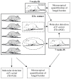
Figure 4. Nude rat model of aerial P. carinii transmission.
To study the airborne transmission of P. carinii, three groups of animals were infected with either P. carinii total population or trophic or cystic forms. To ease comprehension, only a portion (1/4) of the animals belonging to a single group is represented on the figure. First, seeder rats (in grey) were endotracheally infected with P. carinii organisms (total sorted P. carinii population, pure trophic forms, or cystic forms) [23]. Second, after 15 min of recovery, seeder rats were placed in close contact with receiver animals (in white) for 12 h at a mean ratio (seeders to receivers) of 2.4 per cage. These dexamethasone-treated animals were co-housed in capped cages within an individual compartment (thick black square line) of an HEPA-filtered air isolator. Third, all the seeder and half of the receiver rats were euthanized at the end of the contact period. P. carinii organisms were extracted from the seeder rat lungs [29]: the fungal burden was microscopically quantified [26-28] and molecular detection of the P. carinii mtLSUrRNA gene was performed using a single-round touchdown PCR (TD-PCR, [32]). Whole lung tissue suspensions of half of the receiver rats (dotted square line) were screened by nested-PCR (nPCR) at the same locus to detect P. carinii gDNA [5]. Fourth, the other half of the receiver rats (dotted oval line) were kept under immunosuppression (IS) in HEPA-filtered air conditions for 7 weeks to microscopically monitor for eventual PcP development. nPCR was also performed on whole lung tissue suspensions to detect P. carinii gDNA. Whole lung tissue suspensions of sentinel rats were also screened by nPCR and by microscopy after 7 weeks of immunosuppression.

References

    1. Calderón EJ, Gutiérrez-Rivero S, Durand-Joly I, Dei-Cas E (2010) Pneumocystis infection in humans: diagnosis and treatment. Expert Rev Anti Infect Ther 8: 683-701. doi:10.1586/eri.10.42. PubMed: 20521896. - DOI - PubMed
    1. Catherinot E, Lanternier F, Bougnoux ME, Lecuit M, Couderc LJ et al. (2010) Pneumocystis jirovecii Pneumonia. Infect Dis Clin North Am 24: 107-138. doi:10.1016/j.idc.2009.10.010. PubMed: 20171548. - DOI - PubMed
    1. Aliouat-Denis CM, Chabé M, Demanche C, Aliouat EM, Viscogliosi E et al. (2008) Pneumocystis species, co-evolution and pathogenic power. Infect Genet Evol 8: 708-726. doi:10.1016/j.meegid.2008.05.001. PubMed: 18565802. - DOI - PubMed
    1. An CL, Gigliotti F, Harmsen AG (2003) Exposure of immunocompetent adult mice to Pneumocystis carinii f. sp. muris by cohousing: growth of P. carinii f. sp. muris and host immune response. Infect Immun 71: 2065-2070. doi:10.1128/IAI.71.4.2065-2070.2003. PubMed: 12654827. - DOI - PMC - PubMed
    1. Chabé M, Dei-Cas E, Creusy C, Fleurisse L, Respaldiza N et al. (2004) Immunocompetent hosts as a reservoir of Pneumocystis organisms: histological and RT-PCR data demonstrate active replication. Eur J Clin Microbiol Infect Dis 23: 89-97. doi:10.1007/s10096-003-1092-2. PubMed: 14712369. - DOI - PubMed

Publication types