

Evaluation of the sensitivity and specificity of immunohistochemical markers in the differential diagnosis of effusion cytology
- PMID: 24223244
- PMCID: PMC3815850
- DOI: 10.5001/omj.2013.117
Evaluation of the sensitivity and specificity of immunohistochemical markers in the differential diagnosis of effusion cytology
Abstract
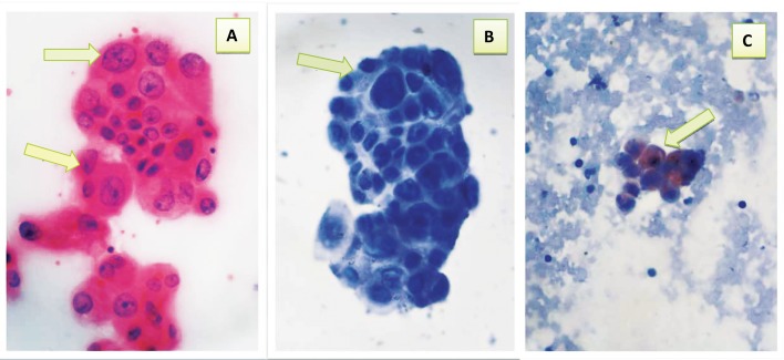
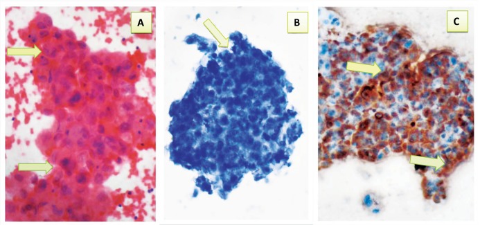
Objective: To evaluate the sensitivity and specificity of Calretinin and Carcinoembryonic antigen as immunocytochemical markers in distinguishing mesothelial cells from metastatic adenocarcinoma cells in effusion cytology.
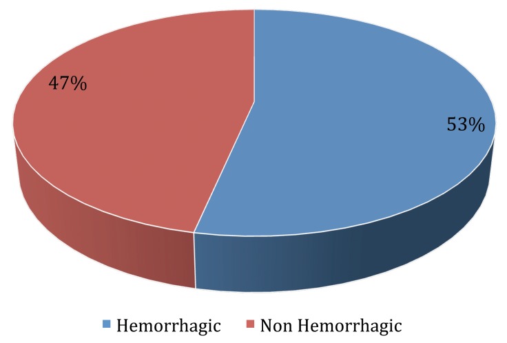
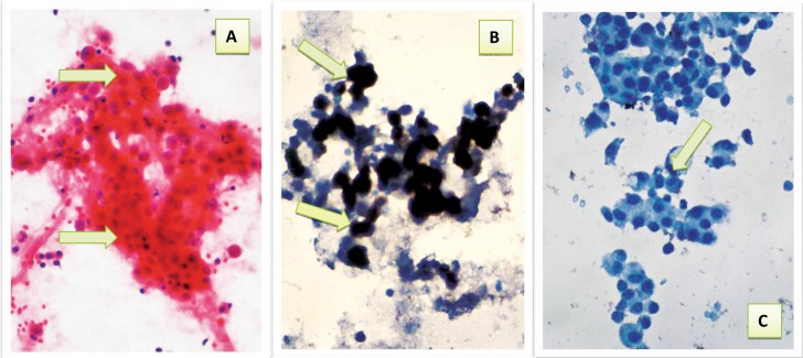
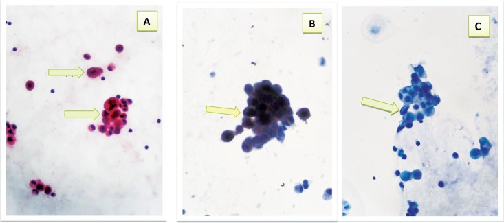
Methods: This study included 50 patients who presented with effusions (26 pleural and 24 peritoneal), at Al-Kadhimya Teaching Hospital who were selected according to their preliminary diagnosis from 1st December 2010 to 30th June 2011. Effusion fluids were aspirated and processed for both conventional cytological methods using Papanicolaou-stain and immunocytochemical staining with anti Calretinin and Carcinoembryonic antigen.
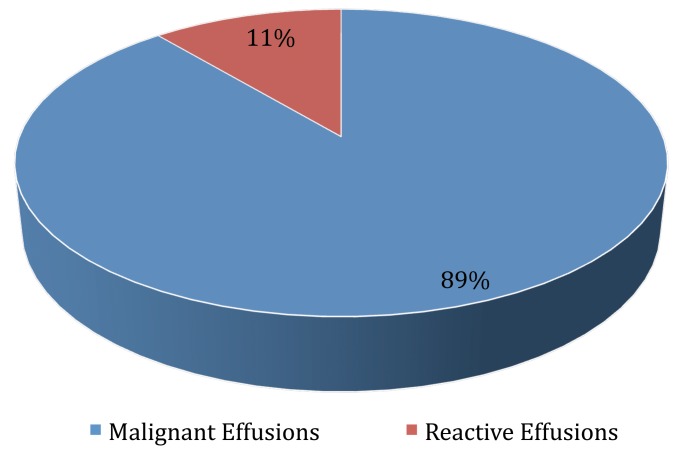
Results: The sensitivity of cytology for detection of malignant cells was 77%, with 100% specificity and 86% accuracy. Calretinin was observed to be a specific (100%) and sensitive (90%) marker for mesothelial cells (of benign etiology). Carcinoembryonic antigen exhibited 70% sensitivity and 100% specificity for adenocarcinoma cells. When the results of both cytology and immunocytochemistry were considered in conjunction, the sensitivity for the detection of malignancy increased to 97%, with 100% specificity and 98% accuracy.
Conclusion: Calretinin and Carcinoembryonic antigen were found to be useful markers for differentiating reactive mesothelial cells from metastatic adenocarcinoma cells in smears prepared from body fluids. Also, the combination of both cytology and immunocytochemical studies using the two markers can greatly enhance the diagnostic accuracy, sensitivity and specificity in malignant effusions.
Keywords: Adenocarcinoma; Calretinin; Carcinoembryonic antigen; Effusion; Mesothelial Cells.
Figures
References
-
- Shidham VB, Falzon M. Serous effusions. In: Gray W, Kocjan G: editors. Diagnostic Cytopathology, 3rd Edition. Churchill Livingstone, Elsevier 2010; p 115-175.
-
- Lyons-Boudreaux V, Mody DR, Zhai J, Coffey D. Cytologic malignancy versus benignancy: how useful are the "newer" markers in body fluid cytology? Arch Pathol Lab Med 2008. Jan;132(1):23-28 - PubMed
-
- Murugan P, Siddaraju N, Habeebullah S, Basu D. Immunohistochemical distinction between mesothelial and adenocarcinoma cells in serous effusions: a combination panel-based approach with a brief review of the literature. Indian J Pathol Microbiol 2009. Apr-Jun;52(2):175-181 10.4103/0377-4929.48910 - DOI - PubMed
LinkOut - more resources
Full Text Sources
Other Literature Sources