. 2013 Nov 4;8(11):e78464.
doi: 10.1371/journal.pone.0078464. eCollection 2013.

Mesenchymal stem cells (MSC) prevented the progression of renovascular hypertension, improved renal function and architecture

Affiliations

Mesenchymal stem cells (MSC) prevented the progression of renovascular hypertension, improved renal function and architecture

Elizabeth B Oliveira-Sales et al. PLoS One. .

Abstract

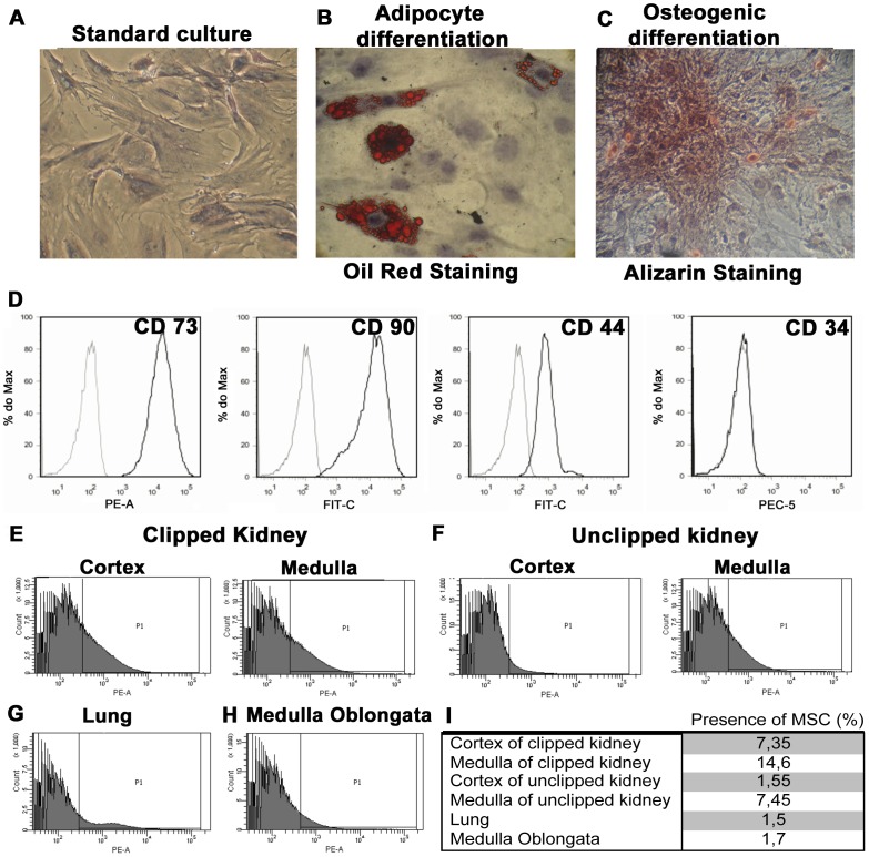
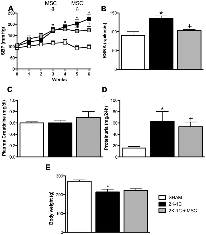
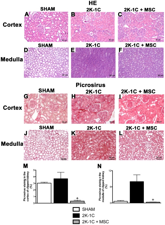
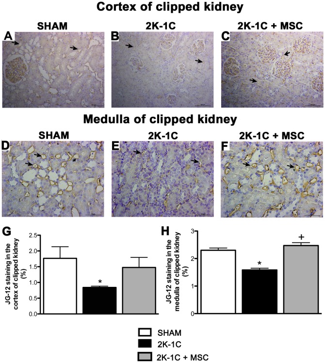
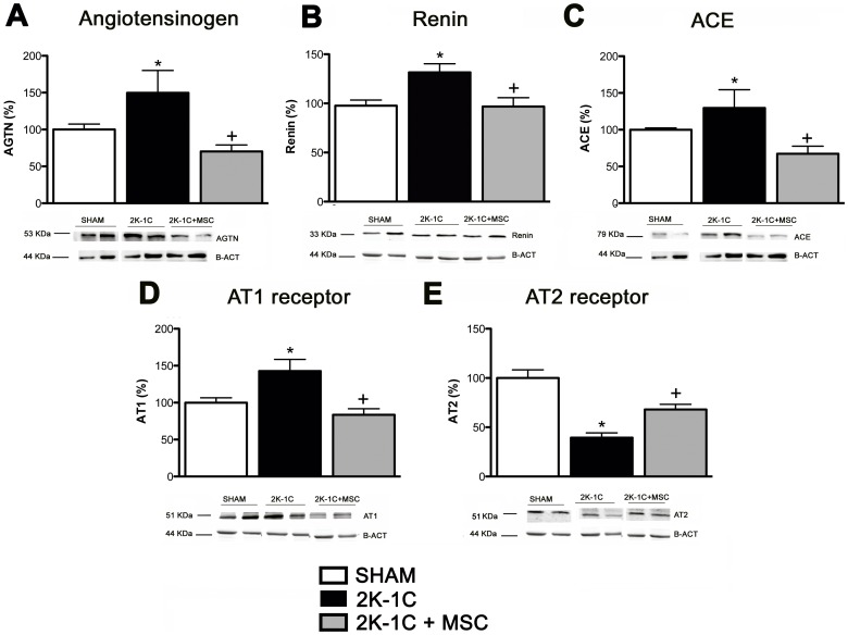
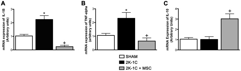
Renovascular hypertension induced by 2 Kidney-1 Clip (2K-1C) is a renin-angiotensin-system (RAS)-dependent model, leading to renal vascular rarefaction and renal failure. RAS inhibitors are not able to reduce arterial pressure (AP) and/or preserve the renal function, and thus, alternative therapies are needed. Three weeks after left renal artery occlusion, fluorescently tagged mesenchymal stem cells (MSC) (2×10(5) cells/animal) were injected weekly into the tail vein in 2K-1C hypertensive rats. Flow cytometry showed labeled MSC in the cortex and medulla of the clipped kidney. MSC prevented a further increase in the AP, significantly reduced proteinuria and decreased sympathetic hyperactivity in 2K-1C rats. Renal function parameters were unchanged, except for an increase in urinary volume observed in 2K-1C rats, which was not corrected by MSC. The treatment improved the morphology and decreased the fibrotic areas in the clipped kidney and also significantly reduced renal vascular rarefaction typical of 2K-1C model. Expression levels of IL-1β, TNF-α angiotensinogen, ACE, and Ang II receptor AT1 were elevated, whereas AT2 levels were decreased in the medulla of the clipped kidney. MSC normalized these expression levels. In conclusion, MSC therapy in the 2K-1C model (i) prevented the progressive increase of AP, (ii) improved renal morphology and microvascular rarefaction, (iii) reduced fibrosis, proteinuria and inflammatory cytokines, (iv) suppressed the intrarenal RAS, iv) decreased sympathetic hyperactivity in anesthetized animals and v) MSC were detected at the CNS suggesting that the cells crossed the blood-brain barrier. This therapy may be a promising strategy to treat renovascular hypertension and its renal consequences in the near future.


Conflict of interest statement

Competing Interests: Dr. Niels Olsen Saraiva Camara, co-author of this article, is member of the PLOS ONE Editorial Board. This does not alter the authors' adherence to all the PLOS ONE policies on sharing data and materials.

Figures

Figure 1
Figure 1. Adipocyte and osteogenic differentiation and MSC tracking assay.
Standard culture (A), differentiated adipocytes containing lipid droplets visualized by Oil Red coloration (B), and osteocytes exhibiting calcium deposition (C), demonstrated by Alizarin coloration. Magnification: 100x. (D) MSC immunophenotyping. MSCs were isolated and labeled for CD73, CD90, CD44 and CD34. Labeled MSC with fluorescent marker detected by cytometry of 2K-1C animals (n = 2) and 2K-1C + MSC (n = 2) in the cortex and medulla of the clipped kidney (E), unclipped kidney (F), lung (G) and medulla oblongata (H). Percentage of labeled cells found in the cortex and medulla of clipped and unclipped kidney, lung and medulla oblongata, analyzed by FACS (I).
Figure 2
Figure 2. Systolic blood pressure (SBP), renal sympathetic nerve activity (RSNA) and renal functional parameters.
(A) SBP expressed as the mean ± SEM. *P<0.05 vs. SHAM. + P<0.05 vs. 2K-1C (2-way ANOVA followed by the Bonferroni’s post-test). RSNA (B), plasma creatinine (C), proteinuria (D) and body weight (E) of SHAM animals (n = 5), 2K-1C animals (n = 7) and 2K-1C + MSC animals (n = 7). *P<0.05 vs. SHAM. + P<0.05 vs. 2K-1C. (One-way ANOVA followed by the Newman Keuls’s post-test).
Figure 3
Figure 3. Morphology and fibrosis of the kidney.
Hematoxylin-Eosin (H&E) (A-F) and Picrosirius red (G-L) staining (20x) of a renal cortex and medulla of the clipped kidneys of SHAM, 2K-1C and 2K-1C + MSC animals. Fibrotic area was measured using the Image Tool program (M and N). * P<0.05 vs. 2K-1C. (One-way ANOVA followed by the Newman Keuls’s post-test.
Figure 4
Figure 4. Capillary staining.
Representative image of JG-12 staining of a renal cortex (20x) and medulla (40X) of the clipped kidney of a SHAM (A and D), 2K-1C (B and E) and 2K-1C + MSC animal (C and F). Stained areas were measured using the Image Tool program (G and H). The arrows show the positive staining in the arterioles. *P<0.05 vs. SHAM. + P<0.05 vs. 2K-1C (One-way ANOVA followed by the Tukey’s post-test).
Figure 5
Figure 5. Expression of components of Renin-Angiotensin (RAS) in the medulla of clipped kidneys.
Angiotensinogen (A), Renin (B), ACE (C), AT1 (D) and AT2 receptors (E) in the medulla of kidney clipped of SHAM (n = 5), 2K-1C (n = 7) and 2K-1C + MSC animals (n = 7). *P<0.05 vs. SHAM. + P<0.05 vs. 2K-1C (One-way ANOVA followed by the Tukey’s post-test).
Figure 6
Figure 6. Cytokine mRNA expression.
(A) IL-1β mRNA expression. (B) TNF-α mRNA expression. (C) IL-10 mRNA expression. Data expressed as arbitrary units *P<0.05 vs. SHAM. + P<0.05 vs. 2K-1C. (One-way ANOVA followed by the Tukey’s post-test).

Similar articles

Cited by

References

    1. Chade AR, Rodriguez-Porcel M, Grande JP, Krier JD, Lerman A, et al. (2002) Distinct renal injury in early atherosclerosis and renovascular disease. Circulation 106: 1165–1171. - PubMed
    1. Chade AR, Rodriguez-Porcel M, Grande JP, Zhu X, Sica V, et al. (2003) Mechanisms of renal structural alterations in combined hypercholesterolemia and renal artery stenosis. Arterioscler Thromb Vasc Biol 23: 1295–1301. - PubMed
    1. Chade AR, Zhu X, Mushin OP, Napoli C, Lerman A, et al.. (2006) Simvastatin promotes angiogenesis and prevents microvascular remodeling in chronic renal ischemia. Faseb J 20: 1706 –1708. - PubMed
    1. Goldblatt H, Lynch J, Hanzal RF, Summerville WW (1934) Studies on experimental hypertension. The production of persistent elevation systolic blood pressure by means of renal ischemia. J Exp Med 59: 347–378. - PMC - PubMed
    1. Ploth DW (1983) Angiotensin-dependent renal mechanisms in two-kidney one-clip renal vascular hypertension. Am J Physiol 245: 131–141. - PubMed

Publication types

MeSH terms