

Chemokine receptors CCR6 and CXCR3 are necessary for CD4(+) T cell mediated ocular surface disease in experimental dry eye disease
- PMID: 24223818
- PMCID: PMC3817213
- DOI: 10.1371/journal.pone.0078508
Chemokine receptors CCR6 and CXCR3 are necessary for CD4(+) T cell mediated ocular surface disease in experimental dry eye disease
Abstract
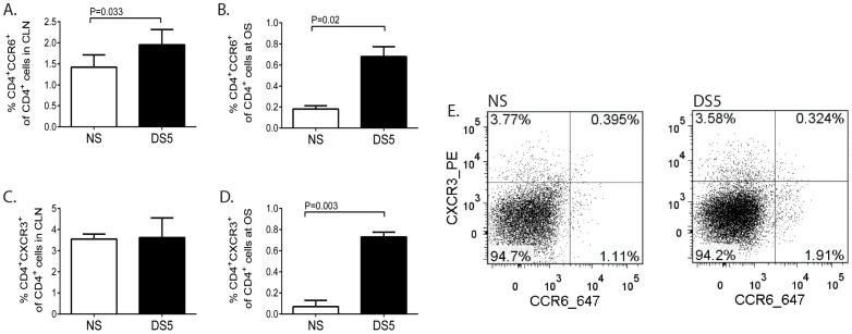
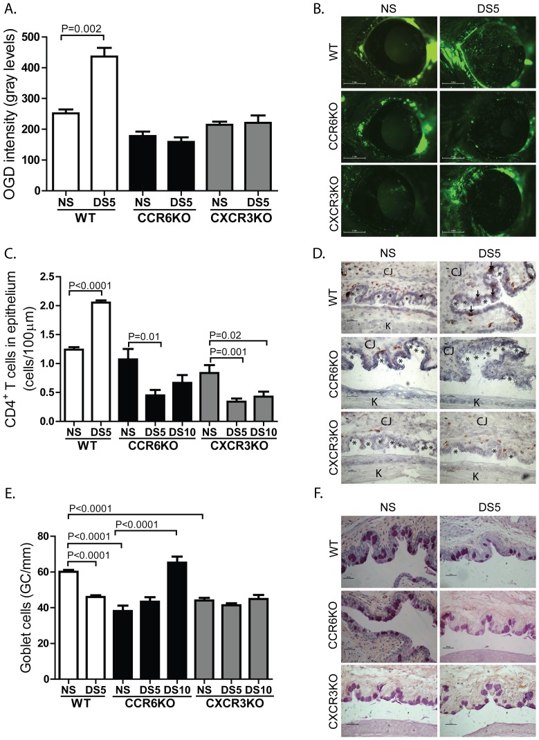
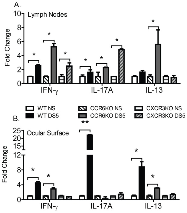
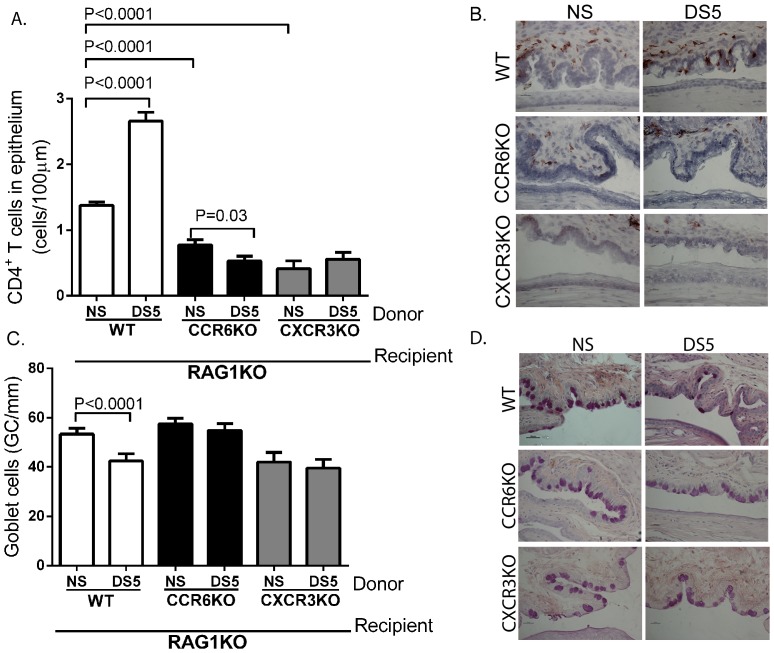
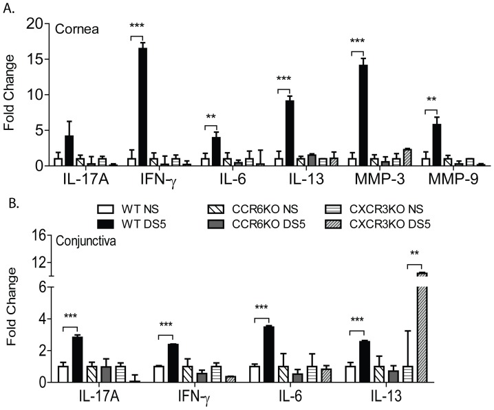
CD4(+) T cells are essential to pathogenesis of ocular surface disease in dry eye. Two subtypes of CD4(+) T cells, Th1 and Th17 cells, function concurrently in dry eye to mediate disease. This occurs in spite of the cross-regulation of IFN-γ and IL-17A, the prototypical cytokines Th1 and Th17 cells, respectively. Essential to an effective immune response are chemokines that direct and summon lymphocytes to specific tissues. T cell trafficking has been extensively studied in other models, but this is the first study to examine the role of chemokine receptors in ocular immune responses. Here, we demonstrate that the chemokine receptors, CCR6 and CXCR3, which are expressed on Th17 and Th1 cells, respectively, are required for the pathogenesis of dry eye disease, as CCR6KO and CXCR3KO mice do not develop disease under desiccating stress. CD4(+) T cells from CCR6KO and CXCR3KO mice exposed to desiccating stress (DS) do not migrate to the ocular surface, but remain in the superficial cervical lymph nodes. In agreement with this, CD4(+) T cells from CCR6 and CXCR3 deficient donors exposed to DS, when adoptively transferred to T cell deficient recipients manifest minimal signs of dry eye disease, including significantly less T cell infiltration, goblet cell loss, and expression of inflammatory cytokine and matrix metalloproteinase expression compared to wild-type donors. These findings highlight the important interaction of chemokine receptors on T cells and chemokine ligand expression on epithelial cells of the cornea and conjunctiva in dry eye pathogenesis and reveal potential new therapeutic targets for dry eye disease.
Conflict of interest statement
Figures
References
-
- Schein OD, Munoz B, Tielsch JM, Bandeen-Roche K, West S (1997) Prevalence of dry eye among the elderly. Am J Ophthalmol 124: 723–728. - PubMed
-
- McCarty CA, Bansal AK, Livingston PM, Stanislavsky YL, Taylor HR (1998) The epidemiology of dry eye in Melbourne, Australia. Ophthalmology 105: 1114–1119. - PubMed
-
- Shimmura S, Shimazaki J, Tsubota K (1999) Results of a population-based questionnaire on the symptoms and lifestyles associated with dry eye. Cornea 18: 408–411. - PubMed
-
- Moss SE, Klein R, Klein BE (2000) Prevalence of and risk factors for dry eye syndrome. Arch Ophthalmol 118: 1264–1268. - PubMed
-
- Moss SE, Klein R, Klein BE (2004) Incidence of dry eye in an older population. Arch Ophthalmol 122: 369–373. - PubMed
Publication types
MeSH terms
Substances
Grants and funding
- 2 T32 EY 7001-36/EY/NEI NIH HHS/United States
- EY018888/EY/NEI NIH HHS/United States
- EY020799/EY/NEI NIH HHS/United States
- S10RR024574/RR/NCRR NIH HHS/United States
- P30CA125123/CA/NCI NIH HHS/United States
- T32 EY007001/EY/NEI NIH HHS/United States
- P30 AI036211/AI/NIAID NIH HHS/United States
- P30 EY002520/EY/NEI NIH HHS/United States
- R21 EY018888/EY/NEI NIH HHS/United States
- S10 RR024574/RR/NCRR NIH HHS/United States
- EY018090/EY/NEI NIH HHS/United States
- EY002520/EY/NEI NIH HHS/United States
- EY11915/EY/NEI NIH HHS/United States
- P30AI036211/AI/NIAID NIH HHS/United States
- P30 CA125123/CA/NCI NIH HHS/United States
- P30 EY020799/EY/NEI NIH HHS/United States
- R01 EY018090/EY/NEI NIH HHS/United States
- R01 EY011915/EY/NEI NIH HHS/United States
LinkOut - more resources
Full Text Sources
Other Literature Sources
Molecular Biology Databases
Research Materials
