. 2013 Nov 4;8(11):e78593.
doi: 10.1371/journal.pone.0078593. eCollection 2013.

Non-genomic estrogen regulation of ion transport and airway surface liquid dynamics in cystic fibrosis bronchial epithelium

Affiliations

Non-genomic estrogen regulation of ion transport and airway surface liquid dynamics in cystic fibrosis bronchial epithelium

Vinciane Saint-Criq et al. PLoS One. .

Abstract

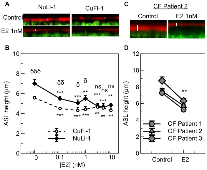
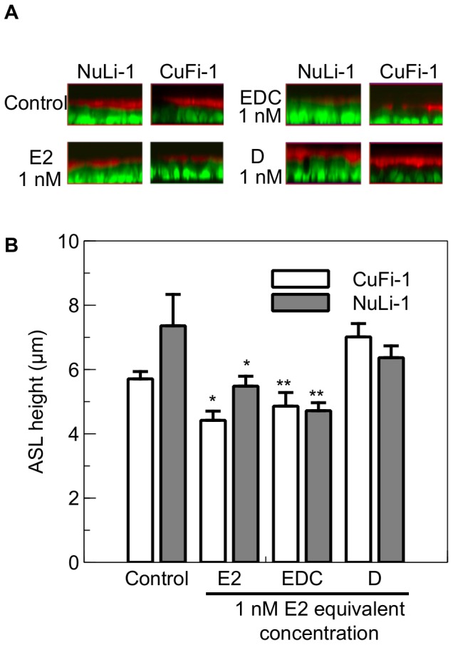
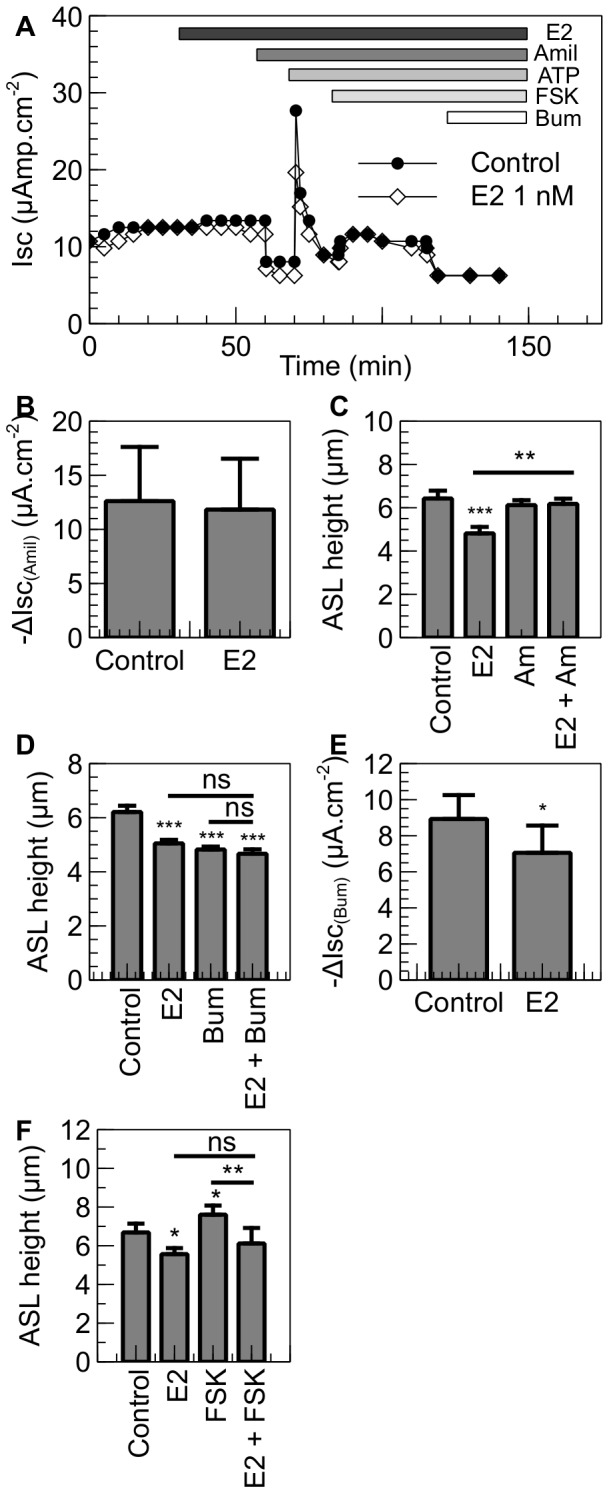
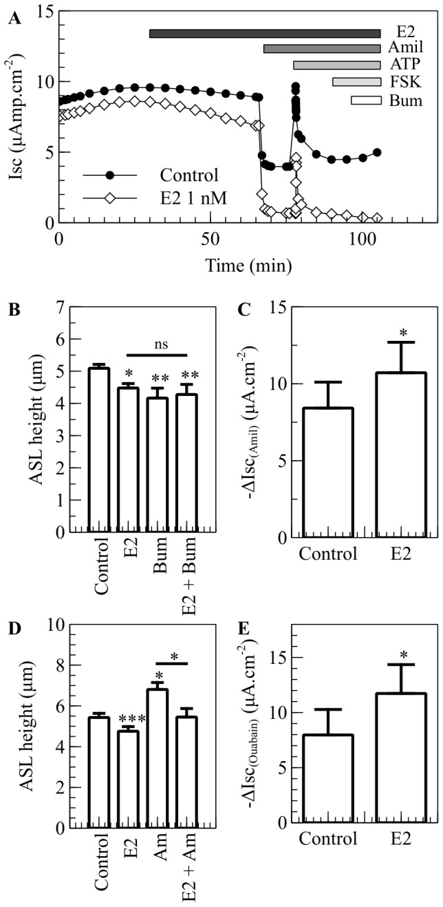
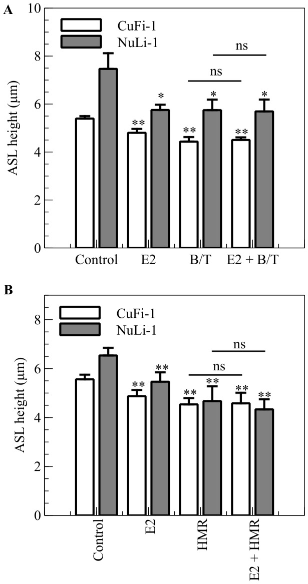
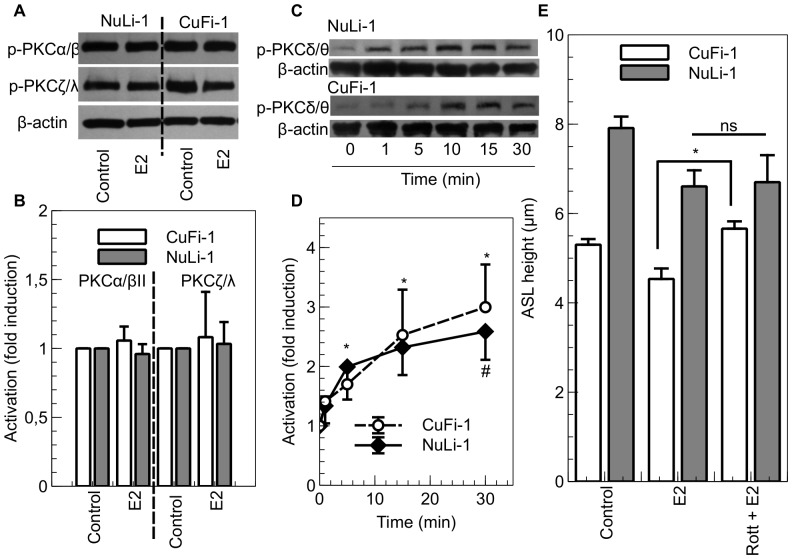
Male cystic fibrosis (CF) patients survive longer than females and lung exacerbations in CF females vary during the estrous cycle. Estrogen has been reported to reduce the height of the airway surface liquid (ASL) in female CF bronchial epithelium. Here we investigated the effect of 17β-estradiol on the airway surface liquid height and ion transport in normal (NuLi-1) and CF (CuFi-1) bronchial epithelial monolayers. Live cell imaging using confocal microscopy revealed that airway surface liquid height was significantly higher in the non-CF cells compared to the CF cells. 17β-estradiol (0.1-10 nM) reduced the airway surface liquid height in non-CF and CF cells after 30 min treatment. Treatment with the nuclear-impeded Estrogen Dendrimer Conjugate mimicked the effect of free estrogen by reducing significantly the airway surface liquid height in CF and non-CF cells. Inhibition of chloride transport or basolateral potassium recycling decreased the airway surface liquid height and 17β-estradiol had no additive effect in the presence of these ion transporter inhibitors. 17β-estradiol decreased bumetanide-sensitive transepithelial short-circuit current in non-CF cells and prevented the forskolin-induced increase in ASL height. 17β-estradiol stimulated an amiloride-sensitive transepithelial current and increased ouabain-sensitive basolateral short-circuit current in CF cells. 17β-estradiol increased PKCδ activity in CF and non-CF cells. These results demonstrate that estrogen dehydrates CF and non-CF ASL, and these responses to 17β-estradiol are non-genomic rather than involving the classical nuclear estrogen receptor pathway. 17β-estradiol acts on the airway surface liquid by inhibiting cAMP-mediated chloride secretion in non-CF cells and increasing sodium absorption via the stimulation of PKCδ, ENaC and the Na(+)/K(+)ATPase in CF cells.


Conflict of interest statement

Competing Interests: The authors have declared that no competing interests exist.

Figures

Figure 1
Figure 1. Concentration-dependence of 17β-estradiol effects on ASL height in NuLi-1 and CuFi-1 epithelial monolayers and the effect of 1 nM E2 on ASL height of primary CF bronchial epithelial cells.
Epithelial cells were stained with Calcein Green and the ASL stained using dextran conjugated Texas Red™ fluorochrome 24 h before estrogen treatment. Panel A shows representative z-plane sections of NuLi-1 (left) and CuFi-1 (right) cells under basal conditions (top) and after 30 mins treatment with 1 nM E2 (bottom). Mean changes in ASL height in control conditions or following treatment with different concentrations of estrogen are shown in panel B for NuLi-1 (solid line) and CuFi-1 (dashed line) cells (n≥4, Error bars reflect standard error of the mean, ANOVA, ** p<0.01, *** p<0.001, compared to control conditions; δ p<0.05, δδ p<0.01, δδδ p<0.001, difference between NuLi-1 and CuFi-1 cells at the indicated estrogen concentration, ns: non significant). Representative z-plane sections of CF primary bronchial epithelial cells under basal conditions (left) and after 30 mins treatment with 1 nM E2 (right) (panel C) and mean changes in ASL height in primary bronchial epithelial cells of 3 CF female children (panel D) (n = 3, paired t-test, ** p<0.01)
Figure 2
Figure 2. Effect of the Estrogen Dendrimer Conjugate on ASL height.
NuLi-1 and CuFi-1 epithelial monolayers were treated with 1 nM E2, or EDC at concentrations providing 1 nM estrogen equivalents, or empty dendrimer at the concentration that matched that in EDC at a 1 nM estrogen equivalent. Panel A shows typical z-plane sections in control conditions or after treatment with E2, EDC or the empty dendrimer (D). The concentrations given for the EDC and the empty dendrimer are in equivalent E2 concentrations. Panel B shows the mean changes in ASL height in control conditions or following exposure to E2, EDC or the empty dendrimer (n≥4, Error bars reflect standard error of the mean, ANOVA, * p<0.05, ** p<0.01).
Figure 3
Figure 3. E2 inhibits Cl secretion in NuLi-1 cells.
NuLi-1 cells were cultured on collagen-coated culture inserts. The monolayers were then allowed to reach a transepithelial resistance of 1,000 Ω.cm−2 or above prior the experiment. A. When mounted in the Ussing chambers, the cells were allowed to stabilize and were then treated basolaterally with E2. The changes in short-circuit current were then measured in response to amiloride (10 µM), ATP (100 µM), forskolin (10 µM) and bumetanide (10 µM). Typical traces of Isc in control and E2 treated NuLi-1 cells are shown. B. Summary of the amiloride short circuit current in control and E2 treated cells. Panel C shows ASLh measurement in NuLi-1 in response to estrogen and ENaC inhibition. NuLi-1 monolayers were treated with E2 (1 nM), Amiloride (Am, 300 µM) or pretreated with estrogen and then treated with amiloride (E2 + Am) (n≥3, error bars reflect standard error of the mean, ANOVA, ** p<0.01, *** p<0.001). Panels D and F show ASLh measurement in NuLi-1 epithelial cells after treatment with (D) E2 (30 min, 1 nM), Bumetanide (Bum, 30 min, 10 µM) or pretreated with E2 and then treated with Bumetanide (E2 + Bum) or (F) E2 (30 min, 1 nM), Forskolin (FSK, 30 min, 10 µM) or pretreated with E2 and then treated with Forskolin (E2 + FSK) (n≥5, Error bars reflect standard error of the mean, ANOVA, * p<0.05, ** p<0.01, *** p<0.001). Panel E shows bumetanide-sensitive current in NuLi-1 cells treated with E2 or with vehicle. Bumetanide was added after 30 minutes (n≥5, Error bars reflect standard error of the mean, Student's t test, * p<0.05).
Figure 4
Figure 4. E2 increases Na+ absorption in CuFi-1 cells.
CuFi-1 cells were cultured on collagen-coated culture inserts. The monolayers were allowed to reach a transepithelial resistance of 1,000 Ω.cm−2 or above prior the experiment. A. In the Ussing chambers, the cells were allowed to stabilize and were then treated basolaterally with E2. The changes in short-circuit current were measured in response to amiloride (10 µM), ATP (100 µM), forskolin (10 µM) and bumetanide (10 µM). Typical traces of Isc in vehicle and E2 treated CuFi-1 cells are shown. B. ASLh was measured in CuFi-1 cells in response to estrogen and NKCC1 inhibition. CuFi-1 monolayers were treated with E2 (1 nM), Bumetanide (Bum, 10 µM) or pretreated with estrogen before bumetanide was added (E2 + Bum) (n = 5, error bars reflect standard error of the mean, ANOVA, * p<0.05, ** p<0.01, ns: non significant). C. Amiloride-sensitive current in CuFi-1 cells treated with E2 or vehicle. Amiloride was added after 30 minutes (n = 6, Error bars reflect standard error of the mean, Student's t test, * p<0.05). D. ASLh measurement in CuFi-1 cells after treatment with E2 (30 min, 1 nM), Amiloride (Amil, 20 min, 300 µM) or pretreated with E2 and then treated with Amiloride (E2 + Amil) (n = 7, Error bars reflect standard error of the mean, ANOVA, * p<0.05, *** p<0.001). F. Ouabain–sensitive current in CuFi-1 cells mounted in Ussing chambers and bathed in modified low Na+ Krebs buffer in which NaCl was replaced by NMDG-Cl. After the current stabilised, the apical membrane was permeabilized and the cells were treated with E2 or with vehicle and Ouabain (100 mM) was added to the basolateral chamber after 30 minutes (n = 4, Error bars reflect standard error of the mean, Student's t test, * p<0.05).
Figure 5
Figure 5. E2 inhibits K+ recycling in normal and CF bronchial cell lines.
A. NuLi-1 (grey bars) and CuFi-1 (white bars) epithelial monolayers were stained with Calcein Green and ASL was stained with dextran conjugated to Texas RedTM fluorochrome and ASL height was measured after treatment with E2 (30 min, 1 nM), and a cocktail of BaCl2 and TEA to inhibit potassium channels (B/T 30 min, BaCl2 10 mM, TEA 1 mM) or pretreated with E2 and then treated with the K+ channels cocktail inhibitor (E2 + B/T) (n = 5, Error bars reflect standard error of the mean, ANOVA, * p<0.05, ** p<0.01). B. NuLi-1 (grey bars) and CuFi-1 (white bars) epithelial monolayers were treated with E2 (30 min, 1 nM), and the KCNQ1 channel specific inhibitor HMR1556 (HMR, 30 min, 1 µM) or pretreated with E2 and then treated with HMR1556 (E2 + HMR) (n≥3, Error bars reflect standard error of the mean, ANOVA, ** p<0.01).
Figure 6
Figure 6. E2-induced PKCδ activation is responsible for the ASL height decrease in CuFi-1 cells.
NuLi-1 (grey bars) and CuFi-1 (white bars) cells were treated basolaterally with E2 (1 nM) for 30 mins. After hormone treatment, cells were washed and total protein extracts were prepared in order to measure the level of phosphorylation of PKCα/βII and PKCζ/λ by immunoblot. Representative Western blots are shown in panel A and densitometric quantitation of phospho-PKCα/βII / β-actin and phospho-PKCζ/λ / β-actin are shown in panel B (n = 4, Error bars reflect standard error of the mean, Student’s t-test). B. NuLi-1 and CuFi-1 cells were treated basolaterally with E2 (1 nM) for the indicated times. After treatment, cells were washed and total protein extracts were prepared in order to measure the level of phosphorylation of PKCδ/θ. Representative images are shown in panel C and mean phosphorylation state of PKCδ/θ changes in control conditions, or following treatment with estrogen at different time points, are shown in panel D (n≥4, Error bars reflect standard error of the mean, ANOVA, * p<0.05). E. ASL height was measured in CuFi-1 (white bars) and NuLi-1 (grey bars) monolayers in response to estrogen and PKCδ inhibition. NuLi-1 and CuFi-1 monolayers were treated with E2 (1 nM) or pretreated with rottlerin (5 µM) and then treated with E2 (n = 5, Error bars reflect standard error of the mean, ANOVA, * p<0.05).

Similar articles

Cited by

References

    1. Musselman TM, Zhang Z, Masilamani SM (2010) Differential regulation of the bumetanide-sensitive cotransporter (NKCC2) by ovarian hormones. Steroids 75: 760–765. - PMC - PubMed
    1. Beers KW, Thompson MA, Chini EN, Dousa TP (1996) beta-Estradiol inhibits Na+-P(i) cotransport across renal brush border membranes from ovarectomized rats. Biochem Biophys Res Commun 221: 442–445. - PubMed
    1. Hofmeister MV, Damkier HH, Christensen BM, Olde B, Fredrik Leeb-Lundberg LM, et al. (2012) 17beta-Estradiol induces nongenomic effects in renal intercalated cells through G protein-coupled estrogen receptor 1. Am J Physiol Renal Physiol 302: F358–368. - PubMed
    1. Cao G, van der Wijst J, van der Kemp A, van Zeeland F, Bindels RJ, et al. (2009) Regulation of the epithelial Mg2+ channel TRPM6 by estrogen and the associated repressor protein of estrogen receptor activity (REA). J Biol Chem 284: 14788–14795. - PMC - PubMed
    1. Groenestege WM, Hoenderop JG, van den Heuvel L, Knoers N, Bindels RJ (2006) The epithelial Mg2+ channel transient receptor potential melastatin 6 is regulated by dietary Mg2+ content and estrogens. J Am Soc Nephrol 17: 1035–1043. - PubMed

Publication types

MeSH terms