. 2013 Nov 4;8(11):e78706.
doi: 10.1371/journal.pone.0078706. eCollection 2013.

Mouse pancreas tissue slice culture facilitates long-term studies of exocrine and endocrine cell physiology in situ

Affiliations

Mouse pancreas tissue slice culture facilitates long-term studies of exocrine and endocrine cell physiology in situ

Anja Marciniak et al. PLoS One. .

Abstract

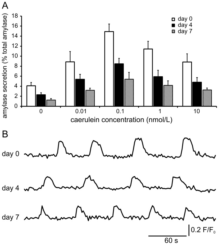
Studies on pancreatic cell physiology rely on the investigation of exocrine and endocrine cells in vitro. Particularly, in the case of the exocrine tissue these studies have suffered from a reduced functional viability of acinar cells in culture. As a result not only investigations on dispersed acinar cells and isolated acini were limited in their potential, but also prolonged studies on pancreatic exocrine and endocrine cells in an intact pancreatic tissue environment were unfeasible. To overcome these limitations, we aimed to establish a pancreas tissue slice culture platform to allow long-term studies on exocrine and endocrine cells in the intact pancreatic environment. Mouse pancreas tissue slice morphology was assessed to determine optimal long-term culture settings for intact pancreatic tissue. Utilizing optimized culture conditions, cell specificity and function of exocrine acinar cells and endocrine beta cells were characterized over a culture period of 7 days. We found pancreas tissue slices cultured under optimized conditions to have intact tissue specific morphology for the entire culture period. Amylase positive intact acini were present at all time points of culture and acinar cells displayed a typical strong cell polarity. Amylase release from pancreas tissue slices decreased during culture, but maintained the characteristic bell-shaped dose-response curve to increasing caerulein concentrations and a ca. 4-fold maximal over basal release. Additionally, endocrine beta cell viability and function was well preserved until the end of the observation period. Our results show that the tissue slice culture platform provides unprecedented maintenance of pancreatic tissue specific morphology and function over a culture period for at least 4 days and in part even up to 1 week. This analytical advancement now allows mid -to long-term studies on the cell biology of pancreatic disorder pathogenesis and therapy in an intact surrounding in situ.


Conflict of interest statement

Competing Interests: The authors have declared that no competing interests exist.

Figures

Figure 1
Figure 1. Preservation of pancreas tissue slice morphology under optimized organotypic culture conditions.
(A) Panels show representative images of pancreas slices immediately after preparation and cultured for 7 days under standard and optimized conditions. Slices cultured in standard conditions exhibit dramatic changes of pancreas morphology whereas slices cultured in optimized conditions preserve the typical lobular structure of dense pancreatic tissue. Scale bars  =  2 mm. (B) Pancreas tissue slice area was maintained significantly better after 4 (88.5±5.3% vs. 59.6±19.2%) and 7 (80.2±6.2% vs. 33.1±15.7%) days when cultured under optimized conditions in comparison to standard conditions. Slice area values are expressed as percent of the area on the day of preparation and represent mean ± SD of n = 12 slices for optimized and n = 64 for standard conditions. (C) Quantification of Draq7 nuclei (dead cells) within exocrine tissue in freshly prepared pancreas tissue slices and during culture in standard and optimized conditions. Exocrine tissue volume was determined by backscatter LSM. Values are expressed as mean ± SD Draq7 labeled nuclei per mm3 exocrine tissue volume (n = 24 for optimized and n = 9 for standard conditions).
Figure 2
Figure 2. Effect of long-term pancreas tissue slice culture under optimized conditions on exocrine tissue morphology and amylase expression.
(A) Immunohistochemistry for amylase (red) and DAPI (blue) in lobules of freshly prepared and under optimized conditions cultured pancreas tissue slices. Labeling revealed dense localization of amylase positive acini at day 0, occupying most of the pancreas lobule area (73.7±7.6%). Amylase labeled acini density and amylase positive area of lobules decreases, however, amylase positive acini are still present at day 4 and 7 of tissue slice culture, (63.9±10.5% and 37.1±7.9%; at day 4 and 7, respectively). Scale bars  = 50 µm. (B) Amylase positive lobule area in fresh and under optimized conditions cultured pancreas tissue slices at indicated time points. Values represent mean ± SD of 16 lobules in 8 slices per time point (n = 16).
Figure 3
Figure 3. Cellular specificity and morphology of exocrine cells in long-term culture of pancreas tissue slices.
(A) Cellular localization of amylase labeling in acini of tissue slices after preparation and on day 4 and 7 of optimized long-term culture. Immunohistochemistry showed polarized localization of amylase labeling at the apical acinar cell pole at all time points. (B) Differential interference contrast (DIC) microscopy of acini in tissue slices at identical time points as in (A). DIC microscopy revealed the presence of zymogen granules at the apical pole and a transparent basal pole of acinar cells in acini of pancreas slices right after preparation and at day 4 and 7 of tissue slice culture. Scale bars  = 10 µm.
Figure 4
Figure 4. Acinar cell function during long-term culture of pancreas tissue slices.
(A) Amylase release from freshly prepared and cultured pancreas tissue slices after 30 min stimulation with indicated caerulein concentrations. The relative amount of released amylase decreases with culture time. However, amylase release shows a typical bell-shaped curve response to increasing caerulein concentrations at all time points. Amylase release is expressed as percent of total amylase as mean ± SD of 16 slices per time point (n = 16). (B) Traces of Ca2+ i/Oregon Green BAPTA-1 fluorescence in acinar cells of pancreas tissue slices at indicated time points after preparation. Stimulation with 10 pmol/L caerulein induced oscillations of Ca2+ i/Oregon Green BAPTA-1 fluorescence in acinar cells of slices at all time points. Traces are shown as fluorescence ratio (F/F0), in which F is fluorescence at any given time and F0 is pre-stimulatory fluorescence.
Figure 5
Figure 5. Effect of long-term pancreas tissue slice culture on endocrine beta cell viability and function.
(A) Longitudinal in situ imaging of beta cell viability and specificity in pancreas tissue slices before and after 7 days culture in standard and optimized conditions. Under standard culture conditions MIP-GFP fluorescence (green) of beta cells was lost after 7 days and the numbers of dead nuclei (magenta) dramatically increased, whereas in optimized conditions beta cells were still detectable by GFP fluorescence after 7 days and the number of dead cells inside the islet remained low. (B) Quantification of Draq7 nuclei (dead cells) within islets in freshly prepared pancreas tissue slices and during culture in standard and optimized conditions. Islet volume in tissue slices was determined by backscatter LSM. Values are expressed as mean ± SD Draq7 labeled nuclei per mm3 islet volume (n = 11 for optimized and n = 12 for standard conditions). (C) Basal (3 mmol/L glucose) and stimulated (16.7 mmol/L glucose) insulin release from pancreas tissue slices at the day of preparation and after 4 and 7 days of culture in optimized conditions. Insulin release is expressed as percent of total insulin as mean ± SD from 8 slices per time point (n = 8).

References

    1. Amsterdam A, Jamieson JD (1974) Studies on dispersed pancreatic exocrine cells. I. Dissociation technique and morphologic characteristics of separated cells. J Cell Biol 63: 1037–1056. - PMC - PubMed
    1. Pipeleers D, in't Veld PI, Maes E, Van De Winkel M (1982) Glucose-induced insulin release depends on functional cooperation between islet cells. Proc Natl Acad Sci U S A 79: 7322–7325. - PMC - PubMed
    1. Bosco D, Soriano JV, Chanson M, Meda P (1994) Heterogeneity and contact-dependent regulation of amylase release by individual acinar cells. J Cell Physiol 160: 378–388. - PubMed
    1. Paraskevas S, Aikin R, Maysinger D, Lakey JR, Cavanagh TJ, et al. (1999) Activation and expression of ERK, JNK, and p38 MAP-kinases in isolated islets of Langerhans: implications for cultured islet survival. FEBS Lett 455: 203–208. - PubMed
    1. Raposo do Amaral AS, Pawlick RL, Rodrigues E, Costal F, Pepper A, et al. (2013) Glutathione Ethyl Ester Supplementation during Pancreatic Islet Isolation Improves Viability and Transplant Outcomes in a Murine Marginal Islet Mass Model. PLoS One 8: e55288. - PMC - PubMed

Publication types

MeSH terms