

Brood ball-mediated transmission of microbiome members in the dung beetle, Onthophagus taurus (Coleoptera: Scarabaeidae)
- PMID: 24223880
- PMCID: PMC3815100
- DOI: 10.1371/journal.pone.0079061
Brood ball-mediated transmission of microbiome members in the dung beetle, Onthophagus taurus (Coleoptera: Scarabaeidae)
Abstract
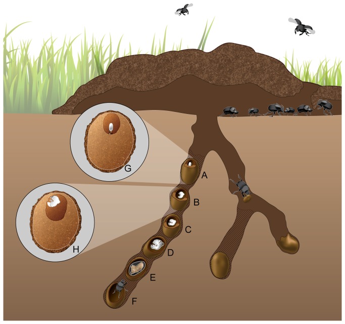
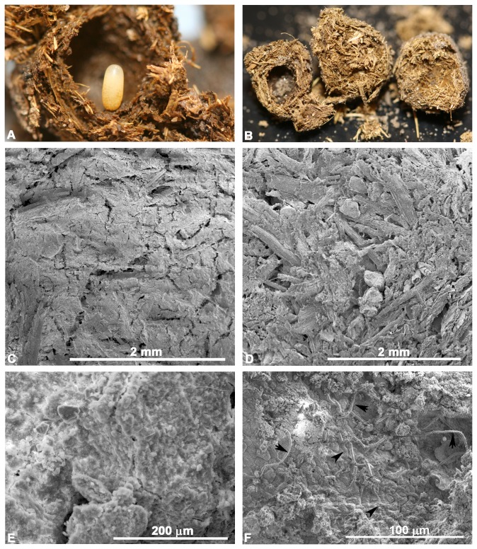
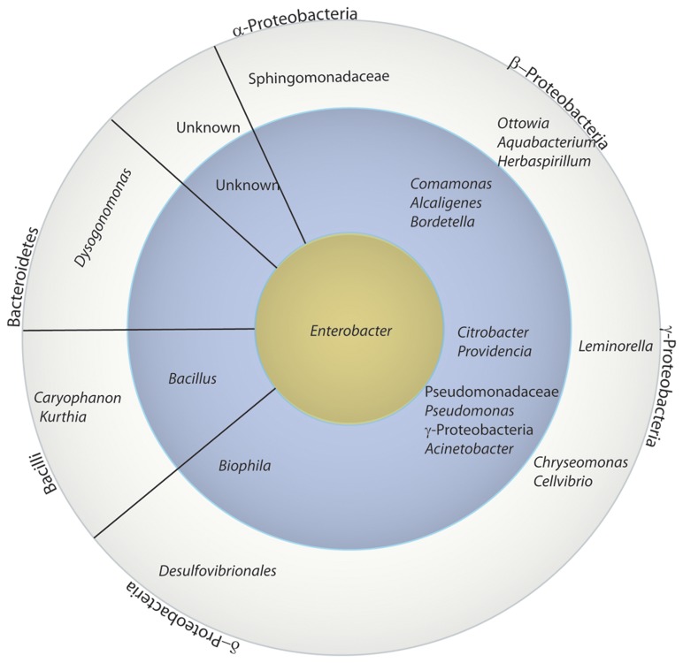
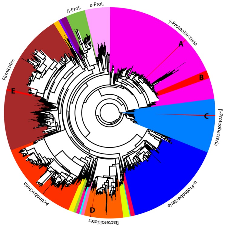
Insects feeding on plant sap, blood, and other nutritionally incomplete diets are typically associated with mutualistic bacteria that supplement missing nutrients. Herbivorous mammal dung contains more than 86% cellulose and lacks amino acids essential for insect development and reproduction. Yet one of the most ecologically necessary and evolutionarily successful groups of beetles, the dung beetles (Scarabaeinae) feeds primarily, or exclusively, on dung. These associations suggest that dung beetles may benefit from mutualistic bacteria that provide nutrients missing from dung. The nesting behaviors of the female parent and the feeding behaviors of the larvae suggest that a microbiome could be vertically transmitted from the parental female to her offspring through the brood ball. Using sterile rearing and a combination of molecular and culture-based techniques, we examine transmission of the microbiome in the bull-headed dung beetle, Onthophagus taurus. Beetles were reared on autoclaved dung and the microbiome was characterized across development. A ~1425 bp region of the 16S rRNA identified Pseudomonadaceae, Enterobacteriaceae, and Comamonadaceae as the most common bacterial families across all life stages and populations, including cultured isolates from the 3(rd) instar digestive system. Finer level phylotyping analyses based on lepA and gyrB amplicons of cultured isolates placed the isolates closest to Enterobacter cloacae, Providencia stuartii, Pusillimonas sp., Pedobacter heparinus, and Lysinibacillus sphaericus. Scanning electron micrographs of brood balls constructed from sterile dung reveals secretions and microbes only in the chamber the female prepares for the egg. The use of autoclaved dung for rearing, the presence of microbes in the brood ball and offspring, and identical 16S rRNA sequences in both parent and offspring suggests that the O. taurus female parent transmits specific microbiome members to her offspring through the brood chamber. The transmission of the dung beetle microbiome highlights the maintenance and likely importance of this newly-characterized bacterial community.
Conflict of interest statement
Figures
References
-
- Chin K, Gill BD (1996) Dinosaurs, dung beetles, and conifers: participants in a Cretaceous food web. Palaios 11: 280-285. doi: 10.2307/3515235. - DOI
-
- Krell F-T (2006) Fossil record and evolution of Scarabaeoidea (Coleoptera: Polyphaga). Coleopt Bull 60: 120-143.
-
- Holter P, Scholtz CH, Stenseng L (2009) Desert detritivory: Nutritional ecology of a dung beetle (Pachysoma glentoni) subsisting on plant litter in arid South African sand dunes. J Arid Environ 73: 1090-1094. doi: 10.1016/j.jaridenv.2009.04.009. - DOI
-
- Davis ALV, Scholtz CH, Philips TK (2002) Historical biogeography of scarabaeine dung beetles. J Biogeogr 29: 1217-1256. doi: 10.1046/j.1365-2699.2002.00776.x. - DOI
Publication types
MeSH terms
Substances
Associated data
- Actions
- Actions
- Actions
- Actions
- Actions
- Actions
- Actions
- Actions
- Actions
- Actions
- Actions
- Actions
- Actions
- Actions
- Actions
- Actions
- Actions
- Actions
- Actions
- Actions
- Actions
- Actions
- Actions
- Actions
- Actions
- Actions
- Actions
- Actions
- Actions
- Actions
- Actions
- Actions
- Actions
- Actions
- Actions
- Actions
- Actions
- Actions
- Actions
- Actions
- Actions
- Actions
- Actions
- Actions
- Actions
- Actions
- Actions
- Actions
- Actions
- Actions
- Actions
- Actions
- Actions
- Actions
- Actions
- Actions
- Actions
- Actions
- Actions
- Actions
- Actions
- Actions
- Actions
- Actions
- Actions
- Actions
- Actions
- Actions
- Actions
- Actions
- Actions
- Actions
- Actions
- Actions
- Actions
- Actions
- Actions
- Actions
- Actions
- Actions
- Actions
- Actions
- Actions
- Actions
- Actions
- Actions
- Actions
- Actions
- Actions
- Actions
- Actions
- Actions
- Actions
- Actions
- Actions
- Actions
- Actions
- Actions
- Actions
- Actions
- Actions
- Actions
- Actions
- Actions
- Actions
- Actions
- Actions
- Actions
- Actions
- Actions
- Actions
- Actions
- Actions
- Actions
- Actions
- Actions
- Actions
- Actions
- Actions
- Actions
- Actions
- Actions
- Actions
- Actions
- Actions
- Actions
- Actions
- Actions
- Actions
- Actions
- Actions
- Actions
- Actions
- Actions
- Actions
- Actions
- Actions
- Actions
- Actions
- Actions
- Actions
- Actions
- Actions
- Actions
- Actions
- Actions
- Actions
- Actions
- Actions
- Actions
- Actions
- Actions
- Actions
- Actions
- Actions
- Actions
- Actions
- Actions
- Actions
- Actions
- Actions
- Actions
- Actions
- Actions
- Actions
- Actions
- Actions
- Actions
- Actions
- Actions
- Actions
- Actions
- Actions
- Actions
- Actions
- Actions
- Actions
- Actions
- Actions
- Actions
- Actions
- Actions
- Actions
- Actions
- Actions
- Actions
- Actions
- Actions
- Actions
- Actions
- Actions
- Actions
- Actions
- Actions
- Actions
- Actions
- Actions
- Actions
- Actions
- Actions
- Actions
- Actions
- Actions
- Actions
- Actions
- Actions
- Actions
- Actions
- Actions
- Actions
- Actions
- Actions
- Actions
- Actions
- Actions
- Actions
- Actions
- Actions
- Actions
- Actions
- Actions
- Actions
- Actions
- Actions
- Actions
- Actions
- Actions
- Actions
- Actions
- Actions
- Actions
- Actions
- Actions
- Actions
- Actions
- Actions
- Actions
- Actions
- Actions
- Actions
- Actions
- Actions
- Actions
- Actions
- Actions
- Actions
- Actions
- Actions
- Actions
- Actions
- Actions
- Actions
- Actions
- Actions
- Actions
- Actions
- Actions
- Actions
- Actions
- Actions
- Actions
- Actions
- Actions
- Actions
- Actions
- Actions
- Actions
- Actions
- Actions
- Actions
- Actions
- Actions
- Actions
- Actions
- Actions
- Actions
- Actions
- Actions
- Actions
- Actions
- Actions
- Actions
- Actions
- Actions
- Actions
- Actions
- Actions
- Actions
- Actions
- Actions
- Actions
- Actions
- Actions
- Actions
- Actions
- Actions
LinkOut - more resources
Full Text Sources
Other Literature Sources
Molecular Biology Databases
Miscellaneous
