Angiotensin II Stimulates Sympathetic Neurotransmission to Adipose Tissue
- PMID: 24224084
- PMCID: PMC3818081
- DOI: 10.1002/phy2.14
Angiotensin II Stimulates Sympathetic Neurotransmission to Adipose Tissue
Abstract
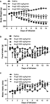
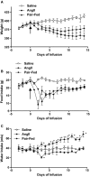
Angiotensin II (AngII) facilitates sympathetic neurotransmission by regulating norepinephrine (NE) synthesis, release and uptake. These effects of AngII contribute to cardiovascular control. Previous studies in our laboratory demonstrated that chronic AngII infusion decreased body weight of rats. We hypothesized that AngII facilitates sympathetic neurotransmission to adipose tissue and may thereby decrease body weight. The effect of chronic AngII infusion on the NE uptake transporter and NE turnover was examined in metabolic (interscapular brown adipose tissue, ISBAT; epididymal fat, EF) and cardiovascular tissues (left ventricle, LV; kidney) of rats. To examine the uptake transporter saturation isotherms were performed using [3H]nisoxetine (NIS). At doses that lowered body weight, AngII significantly increased ISBAT [3H]NIS binding density. To quantify NE turnover, alpha-methyl-para-tyrosine (AMPT) was injected in saline-infused, AngII-infused, or saline-infused rats that were pair-fed to food intake of AngII-infused rats. AngII significantly increased the rate of NE decline in all tissues compared to saline. The rate of NE decline in EF was increased to a similar extent by AngII and by pair-feeding. In rats administered AngII and propranolol, reductions in food and water intake and body weight were eliminated. These data support the hypothesis that AngII facilitates sympathetic neurotransmission to adipose tissue. Increased sympathetic neurotransmission to adipose tissue following AngII exposure is suggested to contribute to reductions in body weight.
Keywords: body weight; catecholamine turnover; norepinephrine.
Figures


Similar articles
-
Facilitation of sympathetic neurotransmission contributes to angiotensin regulation of body weight.J Neural Transm (Vienna). 1999;106(7-8):631-44. doi: 10.1007/s007020050185. J Neural Transm (Vienna). 1999. PMID: 10907723
-
Cold exposure regulates the norepinephrine uptake transporter in rat brown adipose tissue.Am J Physiol. 1999 Jan;276(1):R143-51. doi: 10.1152/ajpregu.1999.276.1.R143. Am J Physiol. 1999. PMID: 9887188
-
Differential effects of local versus systemic angiotensin II in the regulation of leptin release from adipocytes.Endocrinology. 2004 Jan;145(1):169-74. doi: 10.1210/en.2003-0767. Epub 2003 Sep 25. Endocrinology. 2004. PMID: 14512436
-
Angiotensin II in brown adipose tissue from young and adult Zucker obese and lean rats.Am J Physiol. 1994 Mar;266(3 Pt 1):E453-8. doi: 10.1152/ajpendo.1994.266.3.E453. Am J Physiol. 1994. PMID: 8166267
-
Role of AT1 receptors in the central control of sympathetic vasomotor function.Clin Exp Pharmacol Physiol. 1996 Sep;23 Suppl 3:S93-8. doi: 10.1111/j.1440-1681.1996.tb02820.x. Clin Exp Pharmacol Physiol. 1996. PMID: 21143280 Review.
Cited by
-
The inhibitory effect of angiotensin II on BKCa channels in podocytes via oxidative stress.Mol Cell Biochem. 2015 Jan;398(1-2):217-22. doi: 10.1007/s11010-014-2221-1. Epub 2014 Sep 19. Mol Cell Biochem. 2015. PMID: 25234195
-
Angiotensin type 2 receptor activation promotes browning of white adipose tissue and brown adipogenesis.Signal Transduct Target Ther. 2017 Jun 23;2:17022. doi: 10.1038/sigtrans.2017.22. eCollection 2017. Signal Transduct Target Ther. 2017. PMID: 29263921 Free PMC article.
-
Validity of mental and physical stress models.Neurosci Biobehav Rev. 2024 Mar;158:105566. doi: 10.1016/j.neubiorev.2024.105566. Epub 2024 Feb 1. Neurosci Biobehav Rev. 2024. PMID: 38307304 Free PMC article. Review.
-
Potential mechanisms of hypothalamic renin-angiotensin system activation by leptin and DOCA-salt for the control of resting metabolism.Physiol Genomics. 2017 Dec 1;49(12):722-732. doi: 10.1152/physiolgenomics.00087.2017. Epub 2017 Oct 6. Physiol Genomics. 2017. PMID: 28986397 Free PMC article. Review.
-
Classification of Therapeutic and Experimental Drugs for Brown Adipose Tissue Activation: Potential Treatment Strategies for Diabetes and Obesity.Curr Diabetes Rev. 2016;12(4):414-428. doi: 10.2174/1573399812666160517115450. Curr Diabetes Rev. 2016. PMID: 27183844 Free PMC article. Review.
References
-
- Asbert M, Jimenez W, Gaya J, Gines P, Arroyo V, Rivera F, et al. Assessment of the renin-angiotensin system in cirrhotic patients. Comparison between plasma renin activity and direct measurement of immunoreactive renin. J. Hepatol. 1992;15:179–183. - PubMed
-
- Bradford MM. A rapid and sensitive method for the quantitation of microgram quantities of protein utilizing the principle of protein-dye binding. Anal. Biochem. 1976;72:248–254. - PubMed
-
- Brink M, Price SR, Chrast J, Bailey JL, Anwar A, Mitch WE, et al. Angiotensin II induces skeletal muscle wasting through enhanced protein degradation and down-regulates autocrine insulin-like growth factor I. Endocrinology. 2001;142:1489–1496. - PubMed
-
- Brito NA, Brito MN, Bartness TJ. Differential sympathetic drive to adipose tissues after food deprivation, cold exposure or glucoprivation. Am. J. Physiol. Regul. Integr. Comp. Physiol. 2008;294:R1445–R1452. - PubMed
Grants and funding
LinkOut - more resources
Full Text Sources
Other Literature Sources

