. 2013 Jun;2(3):1219.

DNA ligases as therapeutic targets

Affiliations

DNA ligases as therapeutic targets

Alan E Tomkinson et al. Transl Cancer Res. 2013 Jun.

Abstract

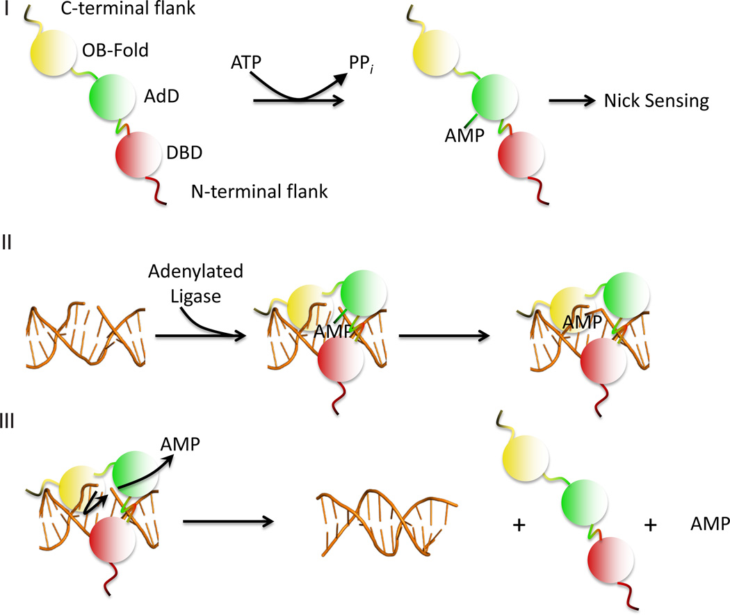
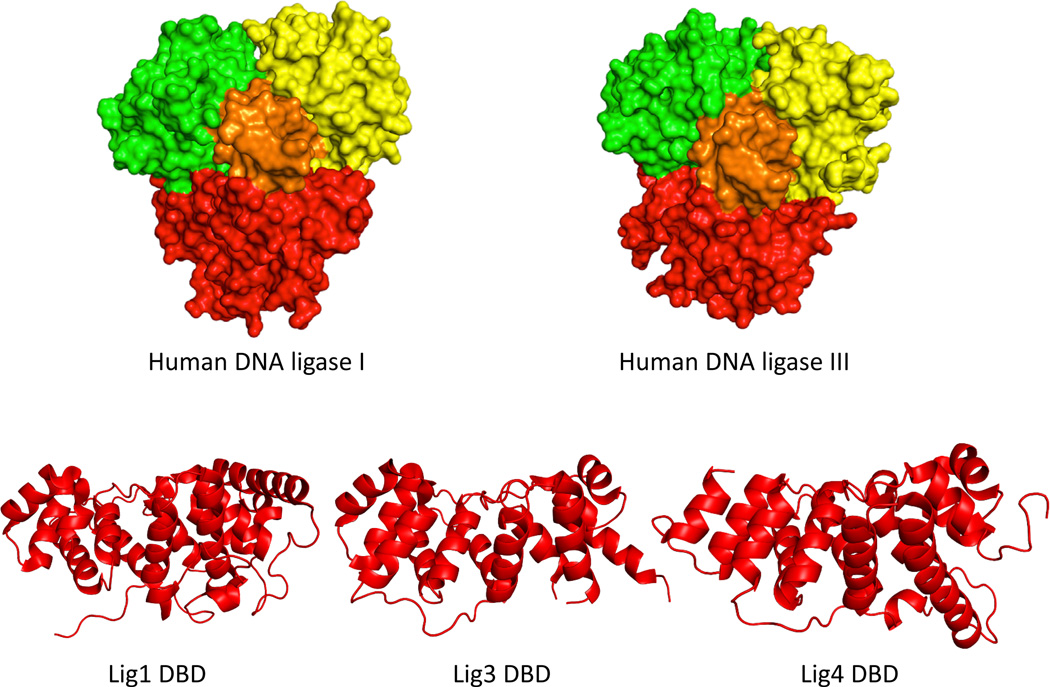
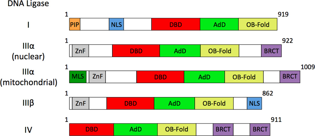
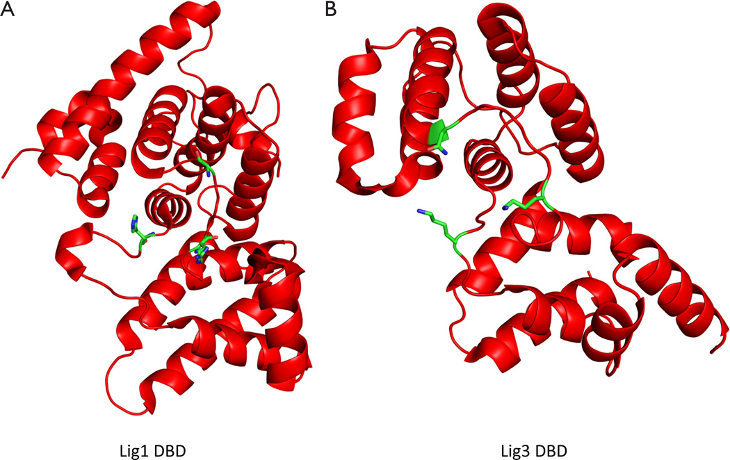
During DNA replication, DNA joining events link Okazaki fragments on the lagging strand. In addition, they are required to repair DNA single- and double-strand breaks and to complete repair events initiated by the excision of mismatched and damaged bases. In human cells, there are three genes encoding DNA ligases. These enzymes are ATP-dependent and contain a conserved catalytic region. Biophysical studies have shown that the catalytic region contains three domains that, in the absence of DNA, are in an extended conformation. When the catalytic region engages a DNA nick, it adopts a compact, ring structure around the DNA nick with each of the three domains contacting the DNA. Protein-protein interactions involving the regions flanking the conserved catalytic regions of human DNA ligases play a major role in directing these enzymes to participate in specific DNA transactions. Among the human LIG genes, the LIG3 gene is unique in that it encodes multiple DNA ligase polypeptides with different N- and C-termini. One of these polypeptides is targeted to mitochondria where it plays an essential role in the maintenance of the mitochondrial genome. In the nucleus, DNA ligases I, III and IV have distinct but overlapping functions in DNA replication and repair. Small molecule inhibitors of human DNA ligases have been identified using structure-based approaches. As expected, these inhibitors are cytotoxic and also potentiate the cytotoxicity of DNA damaging agents. The results of preclinical studies with human cancer cell lines and mouse models of human cancer suggest that DNA ligase inhibitors may have utility as anti-cancer agents.

Keywords: Cancer; DNA ligase; DNA repair; DNA replication; genome instability; mitochondria.


Conflict of interest statement

Disclosure: Dr. Tomkinson is a co-inventor of a patent that covers the use of DNA ligase inhibitors as anticancer agents and a patent application that describes a method for detecting abnormalities in DNA double strand break repair in cancer cells. No potential conflicts of interest were declared by the other authors.

Figures

Figure 1
Figure 1
Steps in the DNA ligation reaction. (I) The catalytic region of the DNA ligase consisting of the DNA binding domain (DBD, red), adenylation domain (AdD, green) and oligonucleotide/oligosaccharide binding-fold (OB-Fold, yellow), interacts with ATP to adenylate an active site lysine within the adenylation domain (AdD, green), releasing pyrophosphate; (II) When the adenylated ligase recognizes and binds to a DNA nick, it undergoes a conformational change such that the DBD, AdD and OB-fold encircle the nick. Within this compact structure, the AMP moiety is transferred from the ligase polypeptide to the 5' phosphate of the nick; (III) The non-adenylated ligase polypeptide utilizes the 3' hydroxyl terminus of the nick as a nucleophile to attack the 5' DNA-adenylate, resulting in phosphodiester bond formation and the release of the ligase polypeptide and AMP
Figure 2
Figure 2
Structures of human DNA ligases. A. Comparison of space-filling representations of the catalytic regions of buman DNA ligase I (Left panel) and human DNA ligase III (Right panel) bound to nicked DNA. The DNA binding domain is indicated in red, the adenylation domain in green, the oligonucleotide/oligosaccharide binding-fold domain in yellow and the nicked DNA in orange; B. Ribbon diagrams showing the secondary structures of the DNA binding domains of human DNA ligases I, III and IV
Figure 3
Figure 3
DNA ligase polypeptides encoded by the human LIG genes. All DNA ligases contain a conserved catalytic region consisting of a DNA-binding domain (DBD, red), an adenylation domain (AdD, green) and an oligosaccharide/oligonucleotide binding-fold (OB-Fold, yellow) domain. The LIG1 gene encodes a single polypeptide with a nuclear localization signal (NLS, blue) and proliferating cell nuclear antigen interacting box (PIP, orange) motif within a non-catalytic N-terminal region. The LIG3 gene encodes multiple polypeptides, each of which contain an N-terminal zinc-finger (ZnF, grey). Mitochondrial and nuclear DNA ligase IIIα are generated by alternative translation initiation with the mitochondrial version having an N-terminal mitochondrial localization signal (MLS, dark green). Both of the DNA ligase IIIα polypeptides have a C-terminal breast and ovarian cancer susceptibility protein 1 C-terminal (BRCT, purple) domain. An alternative splicing event in male germ cells generates DNA ligase IIIβ which has a C-terminal nuclear localization signal (NLS, blue) in place of the BRCT domain. The LIG4 gene encodes a single polypeptide that contains two C-terminal BRCT domains separated by a linker region
Figure 4
Figure 4
DNA binding interface within the DNA binding domain targeted by computer-aided drug design. A. Residues forming the DNA binding pocket within the DNA binding domain (DBD) of human DNA ligase I that was targeted by in silico screening are indicated; B. A similar view of the comparable region within the DBD of human DNA ligase III

References

    1. Friedberg EC, Walker GC, Siede W, et al. DNA Repair and Mutagenesis. 2nd ed. ASM Press; 2006.
    1. Lord CJ, Ashworth A. Targeted therapy for cancer using PARP inhibitors. Curr Opin Pharmacol. 2008;8:363–369. - PubMed
    1. Ellenberger T, Tomkinson AE. Eukaryotic DNA ligases: structural and functional insights. Annu Rev Biochem. 2008;77:313–338. - PMC - PubMed
    1. Pascal JM, O’Brien PJ, Tomkinson AE, et al. Human DNA ligase I completely encircles and partially unwinds nicked DNA. Nature. 2004;432:473–478. - PubMed
    1. Tomkinson AE, Lasko DD, Daly G, et al. Mammalian DNA ligases. Catalytic domain and size of DNA ligase I. J Biol Chem. 1990;265:12611–12617. - PubMed

LinkOut - more resources