Review
. 2014 Jan;10(1):7-19.
doi: 10.4161/auto.26679. Epub 2013 Nov 11.

Boning up on autophagy: the role of autophagy in skeletal biology

Affiliations
Review

Boning up on autophagy: the role of autophagy in skeletal biology

Irving M Shapiro et al. Autophagy. 2014 Jan.

Abstract

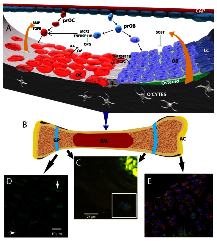
From an evolutionary perspective, the major function of bone is to provide stable sites for muscle attachment and affording protection of vital organs, especially the heart and lungs (ribs) and spinal cord (vertebrae and intervertebral discs). However, bone has a considerable number of other functions: serving as a store for mineral ions, providing a site for blood cell synthesis and participating in a complex system-wide endocrine system. Not surprisingly, bone and cartilage cell homeostasis is tightly controlled, as is the maintenance of tissue structure and mass. While a great deal of new information is accruing concerning skeletal cell homeostasis, one relatively new observation is that the cells of bone (osteoclasts osteoblasts and osteocytes) and cartilage (chondrocytes) exhibit autophagy. The focus of this review is to examine the significance of this process in terms of the functional demands of the skeleton in health and during growth and to provide evidence that dysregulation of the autophagic response is involved in the pathogenesis of diseases of bone (Paget disease of bone) and cartilage (osteoarthritis and the mucopolysaccharidoses). Delineation of molecular changes in the autophagic process is uncovering new approaches for the treatment of diseases that affect the axial and appendicular skeleton.

Keywords: Paget disease of bone; autophagy; bone; cartilage; chondrocytes; growth plate; mucopolysaccharidosis; osteoarthritis; osteoclasts; remodeling; stem cells.


Figures

None
Figure 1. The remodeling process in bone, and autophagy in bone, the growth plate and articular cartilage. (A) Schematic showing molecular control of bone remodeling. A basic multicellular unit consists of osteoblasts (OB) osteoclasts (OC) and bone lining cells (LC). Pro-osteoclasts (PrOC) and pro-osteoblasts (prOB) enter the unit through capillaries (CAP) and home to the bone surface where they undergo differentiation into OB and osteocytes (O’CTES) and OC cells, respectively. Under the influence of local factors including those secreted by osteoblasts (MCF2, TNFSF11), osteoclasts differentiate and resorb bone liberating Ca2+ and amino acids (AA) from the bone matrix. At these sites, a resorption lacuna is formed (RL). Another secreted factor, TNFRSF11B/osteoprotegrin (tumor necrosis factor receptor superfamily, member 11b), inhibits osteoclast-mediated bone resorption by serving as a physiological inhibitor of TNFSF11. A number of factors are released from the resorbing bone that include BMP and TGFB. These proteins promote osteoblast function and maturation. SOST, an inhibitor of osteoblast activity, is also released from the bone matrix. (B) Schematic of a long bone showing articular cartilage (AC), growth plates (GP) and bone marrow (BM). (C) Section through a demineralized osteon showing the presence of autophagic osteocytes (inset, stained with an anti-MAP1LC3 antibody). From Zahm et al., Cells Tissues Organs 2011; 194(2–4):274–8D, and with permission from S Karger AG. (D) Section through the rat growth plate showing the presence of autophagic pre-hypertrophic chondrocytes (arrows) stained with an antibody to MAP1LC3. From Srinivas et al., Cells Tissues Organs. 2009;189(1–4):88–92 and with kind permission from S Karger AG. (E) Chondrocyte autophagy in knee cartilage of GFP-MAP1LC3 transgenic mice. Confocal microscopy-rendered reconstruction using 3D IMARIS (Bitplane Inc.) indicating that the highest levels of GFP-MAP1LC3 signal are observed in chondrocytes in the superficial and upper middle zone of the articular cartilage. In contrast, only a few cells in the deep zone contain detectable levels of GFP-MAP1LC3 signal. Mag × 63

References

    1. Romer AS. The “ancient history” of bone. Ann N Y Acad Sci. 1963;109:168–76. doi: 10.1111/j.1749-6632.1963.tb13466.x. - DOI - PubMed
    1. Karsenty G, Ferron M. The contribution of bone to whole-organism physiology. Nature. 2012;481:314–20. doi: 10.1038/nature10763. - DOI - PMC - PubMed
    1. Boyce BF, Xing L, Shakespeare W, Wang Y, Dalgarno D, Iuliucci J, Sawyer T. Regulation of bone remodeling and emerging breakthrough drugs for osteoporosis and osteolytic bone metastases. Kidney Int Suppl. 2003;85:S2–5. doi: 10.1046/j.1523-1755.63.s85.2.x. - DOI - PubMed
    1. Zhang L, Guo YF, Liu YZ, Liu YJ, Xiong DH, Liu XG, Wang L, Yang TL, Lei SF, Guo Y, et al. Pathway-based genome-wide association analysis identified the importance of regulation-of-autophagy pathway for ultradistal radius BMD. J Bone Miner Res. 2010;25:1572–80. doi: 10.1002/jbmr.36. - DOI - PMC - PubMed
    1. Nagao M, Feinstein TN, Ezura Y, Hayata T, Notomi T, Saita Y, Hanyu R, Hemmi H, Izu Y, Takeda S, et al. Sympathetic control of bone mass regulated by osteopontin. Proc Natl Acad Sci U S A. 2011;108:17767–72. doi: 10.1073/pnas.1109402108. - DOI - PMC - PubMed

Publication types