. 2013 Nov 13;5(211):211ra158.
doi: 10.1126/scitranslmed.3006627.

Sclerostin inhibition reverses skeletal fragility in an Lrp5-deficient mouse model of OPPG syndrome

Affiliations

Sclerostin inhibition reverses skeletal fragility in an Lrp5-deficient mouse model of OPPG syndrome

Rajendra Kedlaya et al. Sci Transl Med. .

Abstract

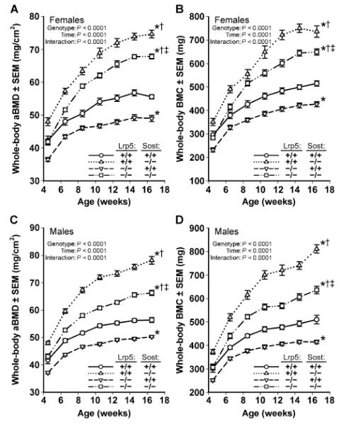
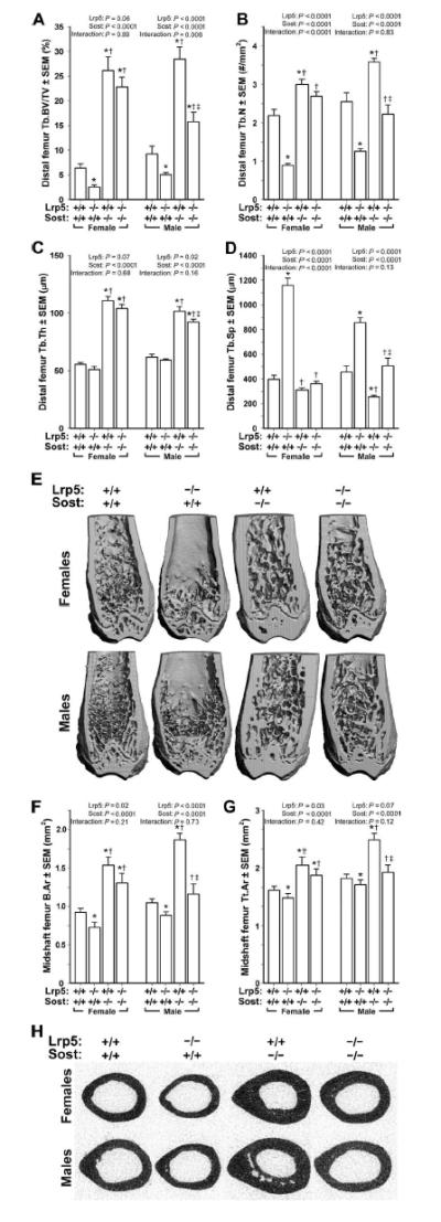
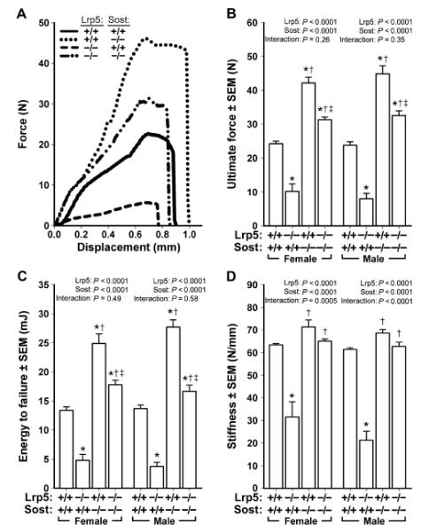
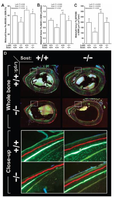
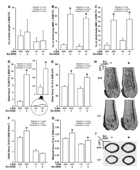
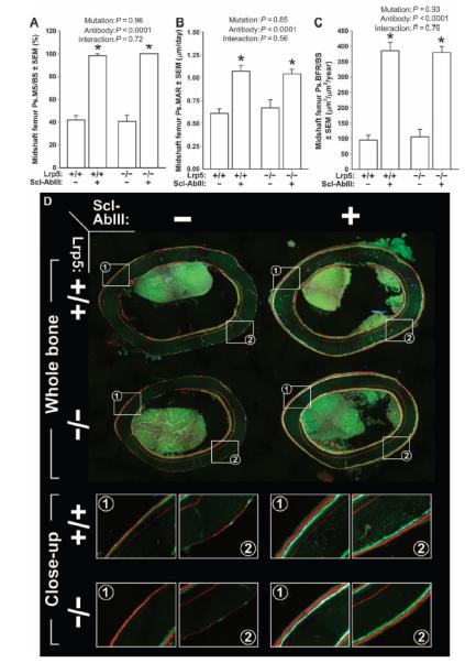
Osteoporosis pseudoglioma syndrome (OPPG) is a rare genetic disease that produces debilitating effects in the skeleton. OPPG is caused by mutations in LRP5, a WNT co-receptor that mediates osteoblast activity. WNT signaling through LRP5, and also through the closely related receptor LRP6, is inhibited by the protein sclerostin (SOST). It is unclear whether OPPG patients might benefit from the anabolic action of sclerostin neutralization therapy (an approach currently being pursued in clinical trials for postmenopausal osteoporosis) in light of their LRP5 deficiency and consequent osteoblast impairment. To assess whether loss of sclerostin is anabolic in OPPG, we measured bone properties in a mouse model of OPPG (Lrp5(-/-)), a mouse model of sclerosteosis (Sost(-/-)), and in mice with both genes knocked out (Lrp5(-/-);Sost(-/-)). Lrp5(-/-);Sost(-/-) mice have larger, denser, and stronger bones than do Lrp5(-/-) mice, indicating that SOST deficiency can improve bone properties via pathways that do not require LRP5. Next, we determined whether the anabolic effects of sclerostin depletion in Lrp5(-/-) mice are retained in adult mice by treating 17-week-old Lrp5(-/-) mice with a sclerostin antibody for 3 weeks. Lrp5(+/+) and Lrp5(-/-) mice each exhibited osteoanabolic responses to antibody therapy, as indicated by increased bone mineral density, content, and formation rates. Collectively, our data show that inhibiting sclerostin can improve bone mass whether LRP5 is present or not. In the absence of LRP5, the anabolic effects of SOST depletion can occur via other receptors (such as LRP4/6). Regardless of the mechanism, our results suggest that humans with OPPG might benefit from sclerostin neutralization therapies.


Figures

Fig. 1
Fig. 1. Longitudinal whole-body dual-energy x-ray absorptiometry (DEXA)–derived measures of areal BMD (aBMD) (A and C) and BMC (B and D), collected in female (A and B) and male (C and D) mice
Scans were collected biweekly beginning at 4.5 weeks of age until 16.5 weeks of age in wild-type mice (circles; solid line), Sost−/− mice (triangles; dotted line), Lrp5−/− mice (inverted triangles; dashed line), and Sost−/− Lrp5−/− double knockouts (squares; interrupted dashed line). *P < 0.05, significant difference from wild-type mice; †P < 0.05, significant difference from Lrp5−/− mice; ‡P < 0.05, significant difference from Sost−/− mice, using repeated-measures analysis of variance (ANOVA). For each group, n = 9 to 14 mice.
Fig. 2
Fig. 2. mCT-derived measurements of the distal femur trabecular bone and midshaft femur cortical bone in wild-type, Lrp5−/−, Sost−/−, and Lrp5−/− Sost−/− mice, collected from male and female mice at 16.5 weeks of age
(A) Trabecular bone volume fraction (BV/TV). (B) Trabecular number (Tb.N). (C) Trabecular thickness (Tb.Th). (D) Trabecular separation (Tb.Sp). (E) Representative cut-away (anterior portion digitally removed to reveal the metaphysis) μCT images of distal femur from the genotypes indicated in (A) to (D). Note the reduced trabecular bone mass induced by the Lrp5 mutation, the marked increase in trabecular bone induced by the Sost mutation, and the elevated trabecular bone mass in the double knockouts. (F and G) Midshaft femur bone tissue area (B.Ar) (F) and midshaft femur total area (Tt.Ar) (G) within the periosteal boundary. (H) Representative midshaft femur μCT images from the genotypes indicated in (F) and (G). Note the reduced cortical bone mass induced by the Lrp5 mutation, the marked increase in cortical bone induced by the Sost mutation, and the elevated cortical bone mass in the double knockouts. The data were analyzed by two-way ANOVA within sex using Lrp5 and Sost genotypes as main effects (indicated at the top of each panel). Post hoc tests were conducted using Fisher’s protected least significant difference (PLSD). *P < 0.05, significantly different from wild type; P < 0.05, significantly different from Lrp5−/−; P < 0.05, significantly different from Sost−/−. For each group, n =9 to 14 mice.
Fig. 3
Fig. 3. Monotonic three-point bending tests to failure of femora from 16.5-week-old male and female wild-type, Lrp5−/−, Sost−/−, and Lrp5−/−;Sost−/− mice
(A) Representative force-displacement curves (male mice depicted) derived from the four genotypes tested. (B to D) Note the mutation-associated changes in peak curve height [ultimate force; quantified in (B)], area under the curve [energy to failure; quantified in (C)], and slope of the elastic portion of the curve [stiffness; quantified in (D)]. The data were analyzed by two-way ANOVA within sex using Lrp5 and Sost genotypes as main effects (indicated at the top of each panel). Post hoc tests were conducted using Fisher’s PLSD. *P < 0.05, significantly different from wild type; P < 0.05, significantly different from Lrp5−/−; P < 0.05, significantly different from Sost−/−. For each group, n = 9 to 14 mice.
Fig. 4
Fig. 4. Midshaft femur fluorochrome-derived BFRs on the periosteal surface collected from 16.5-week-old male Lrp5−/−, Sost−/−, and Lrp5−/−;Sost−/− mice
(A) Periosteal MS/BS (Ps.MS/BS). (B) Periosteal MAR (Ps.MAR). (C) Periosteal BFR per unit bone surface (Ps.BFR/BS). All three indices were derived using an oxytetracycline label given at 5 weeks of age [pale yellow label in (D)] and an alizarin complexone label given at 12 weeks of age [red label in (D)]. (D) Whole-bone (upper panels) and close-up (lower panels; taken from the white boxes indicated in the upper panels) photomicrographs of representative midshaft femur sections from each of the four genotypes studied. The data were analyzed by two-way ANOVA using Lrp5 and Sost genotypes as main effects [indicated at the top of panels (A) to (C)]. Post hoc tests were conducted using Fisher’s PLSD. *P < 0.05, significantly different from wild type; P < 0.05, significantly different from Lrp5−/−; P < 0.05, significantly different from Sost−/−. For each group, n = 8 mice.
Fig. 5
Fig. 5. DEXA- and mCT-derived measurements of bone mass, density, and architecture in female Lrp5+/+ and Lrp5−/− mice that had been treated for 3 weeks with vehicle or sclerostin antibody (Scl-AbIII)
(A) Percent change in body weight over the 3-week experimental period. (B) Percent change in whole-body aBMD over the 3-week experimental period. (C) Percent change in whole-body BMC over the 3-week experimental period. (D) Distal femur trabecular bone volume fraction (BV/TV) after 3 weeks of treatment with vehicle or antibody. An enlarged view of the antibody effect in Lrp5−/− mice is provided (circle with arrow) because baseline BV/TV is so low in these mice. (E) Trabecular thickness (Tb.Th) after 3 weeks of treatment with vehicle or antibody. (F) Trabecular number (Tb.N) after 3 weeks of treatment with vehicle or antibody. (G) Midshaft femur cortical bone area (B.Ar) after 3 weeks of treatment with vehicle or antibody. (H) Representative cut-away (anterior portion digitally removed to reveal the metaphysis) mCT images of distal femur from the treatment groups indicated in (A) to (G). (I) Representative midshaft femur mCT slice from the treatment groups indicated in (A) to (G). The data were analyzed by two-way ANOVA using Lrp5 genotype and antibody/vehicle treatment as main effects (indicated at the top of each data panel). Post hoc tests comparing antibody treatment to vehicle treatment within Lrp5 genotypes were conducted using Fisher’s PLSD. *P < 0.05, significant difference from vehicle-treated mice. For each group, n = 8 mice.
Fig. 6
Fig. 6. Midshaft femur fluorochrome-derived BFRs on the periosteal surface of 20-week-old female Lrp5+/+ and Lrp5−/− mice that had been treated for 3 weeks with vehicle or a sclerostin antibody (Scl-AbIII)
(A) Periosteal MS/BS (Ps.MS/BS). (B) Periosteal MAR (Ps.MAR). (C) Periosteal BFR per unit bone surface (Ps.BFR/BS). All three indices were derived using a calcein label given at 18 weeks of age [green label in (D)] and an alizarin complexone label given at 19 weeks of age [red label in (D)]. (D) Whole-bone (upper panels) and close-up (lower panels; taken from the white boxes indicated in the upper panels) photomicrographs of representative midshaft femur sections from each of the groups studied. A xylenol orange label can be seen in some of the sections buried deeper in the cortex (given at 12 weeks of age), which served as a pretreatment marker and was not used for any of the dynamic measurements. The data were analyzed by two-way ANOVA using Lrp5 genotype and antibody/vehicle treatment as main effects (indicated at the top of each panel). Post hoc tests comparing antibody treatment to vehicle treatment within Lrp5 genotypes were conducted using Fisher’s PLSD. *P < 0.05, significant difference from vehicle-treated mice. For each group, n = 8 mice.

Comment in

References

    1. Becker DJ, Kilgore ML, Morrisey MA. The societal burden of osteoporosis. Curr. Rheumatol. Rep. 2010;12:186–191. - PubMed
    1. Burge R, Dawson-Hughes B, Solomon DH, Wong JB, King A, Tosteson A. Incidence and economic burden of osteoporosis-related fractures in the United States, 2005–2025. J. Bone Miner. Res. 2007;22:465–475. - PubMed
    1. Robbins J, Aragaki AK, Kooperberg C, Watts N, Wactawski-Wende J, Jackson RD, LeBoff MS, Lewis CE, Chen Z, Stefanick ML, Cauley J. Factors associated with 5-year risk of hip fracture in postmenopausal women. JAMA. 2007;298:2389–2398. - PubMed
    1. Gong Y, B. Slee R, Fukai N, Rawadi G, Roman-Roman S, Reginato AM, Wang H, Cundy T, Glorieux FH, Lev D, Zacharin M, Oexle K, Marcelino J, Suwairi W, Heeger S, Sabatakos G, Apte S, Adkins WN, Allgrove J, Arslan-Kirchner M, Batch JA, Beighton P, Black GC, Boles RG, Boon LM, Borrone C, Brunner HG, Carle GF, Dallapiccola B, De Paepe A, Floege B, Halfhide ML, Hall B, Hennekam RC, Hirose T, Jans A, Jüppner H, Kim CA, Keppler-Noreuil K, Kohlschuetter A, LaCombe D, Lambert M, Lemyre E, Letteboer T, Peltonen L, Ramesar RS, Romanengo M, Somer H, Steichen-Gersdorf E, Steinmann B, Sullivan B, Superti-Furga A, Swoboda W, van den Boogaard MJ, Van Hul W, Vikkula M, Votruba M, Zabel B, Garcia T, Baron R, Olsen BR, Warman ML. Osteoporosis-Pseudoglioma Syndrome Collaborative Group, LDL receptor-related protein 5 (LRP5) affects bone accrual and eye development. Cell. 2001;107:513–523. - PubMed
    1. Saarinen A, Saukkonen T, Kivelä T, Lahtinen U, Laine C, Somer M, Toiviainen-Salo S, Cole WG, Lehesjoki AE, Mäkitie O. Low density lipoprotein receptor-related protein 5 (LRP5) mutations and osteoporosis, impaired glucose metabolism and hypercholesterolaemia. Clin. Endocrinol. 2010;72:481–488. - PubMed

Publication types

MeSH terms

Substances

Supplementary concepts