. 2013 Dec 20;288(51):36691-702.
doi: 10.1074/jbc.M113.512806. Epub 2013 Nov 13.

Body fluid exosomes promote secretion of inflammatory cytokines in monocytic cells via Toll-like receptor signaling

Affiliations

Body fluid exosomes promote secretion of inflammatory cytokines in monocytic cells via Toll-like receptor signaling

Niko P Bretz et al. J Biol Chem. .

Abstract

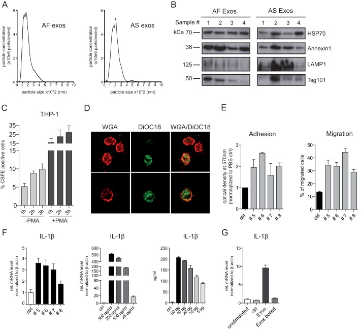
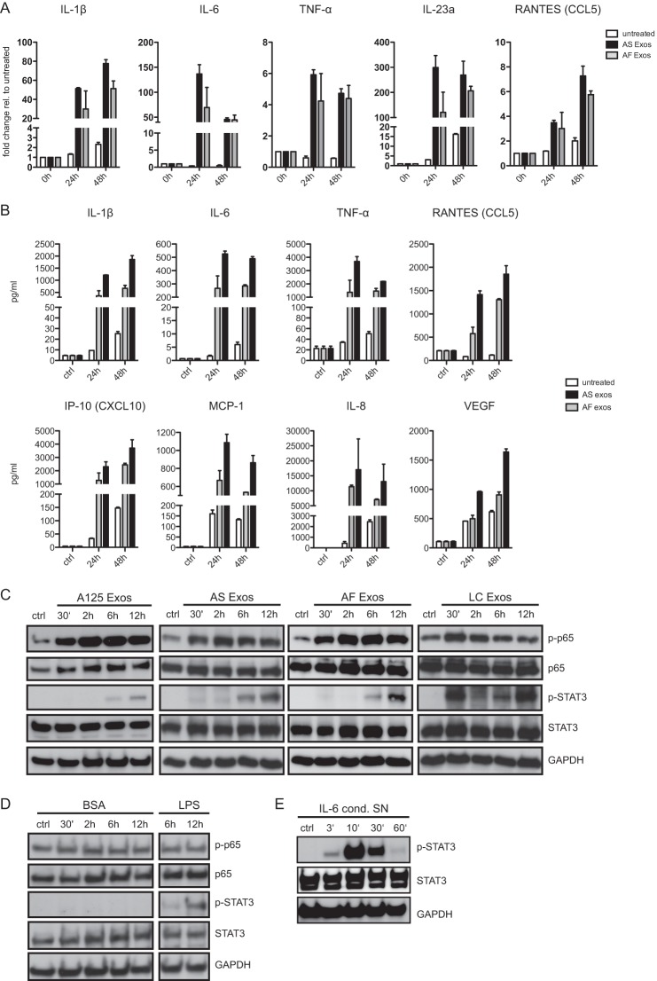
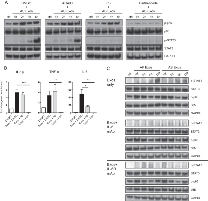
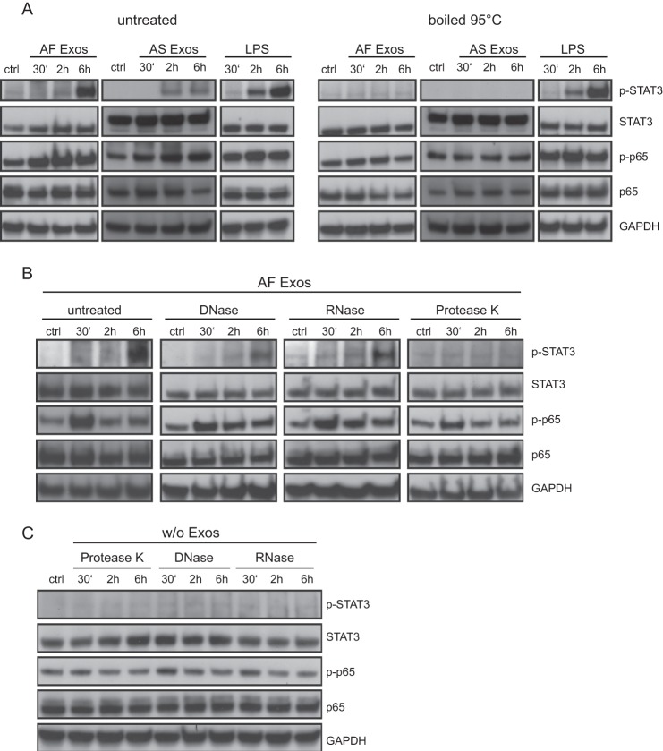
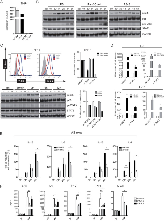
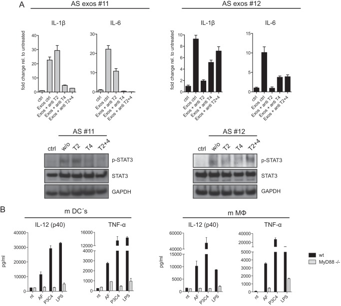
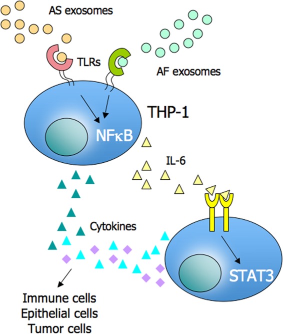
Tumor-derived exosomes have been shown to induce various immunomodulatory effects. However, the underlying signaling pathways are poorly understood. Here, we analyzed the effects of ex vivo-derived exosomes on monocytic cell differentiation/activation using THP-1 cells as model. We isolated exosomes from various body fluids such as amniotic fluid, liver cirrhosis ascites, and malignant ascites of ovarian cancer patients. We observed that exosomes were internalized by THP-1 cells and induced the production of IL-1β, TNF-α, and IL-6. Analysis of the signaling pathways revealed a fast triggering of NFκB and a delayed activation of STAT3. Pharmacologic and antibody-blocking experiments showed that the initial production of IL-6 was instrumental for subsequent activation of STAT3. Importantly, triggering of cell signaling was not a unique property of tumor exosomes but was also observed with exosomes of noncancerous origin. Exosomal signaling was TLR-dependent as the knockdown of Toll-like receptor 2 (TLR2) and TLR4 blocked NFκB and STAT3 activation. Similar results were obtained with TLR-neutralizing antibodies. Exosomes also triggered the release of cytokines from mouse bone marrow-derived dendritic cells or macrophages. This process was MyD88-dependent, further supporting a role of TLR signaling. Our results suggest that exosomes trigger TLR-dependent signaling pathways in monocytic precursor cells but possibly also in other immune cells. This process could be important for the induction of immunosuppressive mechanisms during cancer progression and inflammatory diseases.

Keywords: Amniotic Fluid; Cytokine Induction; Exosomes; Malignant Ascites; NFκB Transcription Factor; STAT3; Toll-like receptor (TLR).


Figures

FIGURE 1.
FIGURE 1.
Characterization of AF and AS exosomes. A, nanosight analysis of isolated AF and AS exosomes is shown. Exosomes were isolated by differential centrifugation from AF or AS of ovarian carcinoma patients. B, representative Western blot analysis of exosomes isolated from four different AF or AS donors is shown. C, AS exosomes were labeled with CFSE and 1 mg of exosomes and exposed to THP-1 cells treated or nontreated before with PMA at 37 °C. After the indicated time point, aliquots of cells were washed and analyzed by cytofluorographic analysis. Mean fluorescence of labeled cells after uptake from three exosomal donors is plotted. D, THP-1 cells that had internalized CFSE exosomes for 1 h were analyzed by fluorescent microscopy. To visualize the plasma membrane the cells were counterstained with phycoerythrin-coupled wheat germ agglutinin (WGA). Note the intracellular localization of labeled exosomes. E, AS exosomes from different donors (#5–8) trigger plastic adhesiveness and cell migration of THP-1 cells. Motility of cells was determined after 24 h by Boyden chamber analysis. F, exosomes induced IL-1β expression. THP-1 cells were incubated with the AS exosomes (10 μg/ml) (donors #5–8) for 24 h at 37 °C. IL-1β mRNA expression was measured by RT-PCR. THP-1 cells were incubated with the indicated amounts of AS exosomes (donor #5) for 24 h at 37 °C. Levels of IL-1β mRNA were measured by RT-PCR, and the cell culture supernatant was analyzed by IL-1β-specific ELISA. G, boiling destroys the stimulating activity. THP-1 cells incubated with 50 μg/ml AS exosomes either boiled (5 min, 95 °C) or nonboiled were analyzed for IL-1β by RT-PCR after 24 h at 37 °C. Error bars, S.E.
FIGURE 2.
FIGURE 2.
Exosomes trigger cytokine production and cell signaling in THP-1 cells. A, cells were incubated with 40 μg/ml AF or AS exosomes for the indicated times at 37 °C and analyzed by RT-PCR for the expression of cytokines. Pooled results of n = 3 experiments are shown. RANTES, regulated on activation normal T cell expressed and secreted. Error bars, S.E. B, exposure of THP-1 cells to exosomes induces secretion of cytokines. Cells were incubated with 40 μg/ml exosomes for the indicated length of time at 37 °C, and the culture supernatant was analyzed by multiplex ELISA. A representative experiment of n = 3 experiments is shown. C, cells were incubated with 40 μg/ml A125, AS, AF, or LC exosomes for the indicated length of time at 37 °C. Cell lysates were analyzed by Western blotting. A representative of n > 3 experiments is shown. D, THP-1 cells were incubated with BSA (40 μg/ml, as negative control) or LPS (1 μg/ml, as positive control) for the indicated length of time. Note that BSA does not activate STAT3 phosphorylation whereas LPS does. E, THP-1 cells were incubated with IL-6 containing cell culture supernatant (SN) for the indicated length of time followed by Western blot analysis.
FIGURE 3.
FIGURE 3.
Role of NFκB and STAT3 in THP-1 cell signaling. A, THP-1 cells were incubated with 40 μg/ml AS exosomes for the indicated length of time at 37 °C in the absence or presence of the JAK2 inhibitor P6 (2 μm), the JAK1 inhibitor AG490 (20 μm), or the specific NFκB inhibitor parthenolide (10 μm). Dimethyl sulfoxide (DMSO) served as solvent control. Cell lysates were prepared and analyzed by Western blotting with the indicated primary antibodies followed by peroxidase-conjugated secondary antibodies and ECL detection. B, RT-PCR analysis was performed on mRNAs isolated from cells treated in A. Error bars, S.E. C, THP-1 cells were incubated with exosomes as described above in the presence or absence of neutralizing antibodies to IL-6 and IL-6 receptor (each 1 μg/ml). Cell lysates were prepared and analyzed by Western blotting as described. ***, p < 0.001; **, p <0.01; *, p <0.05.
FIGURE 4.
FIGURE 4.
Proteinase-sensitive determinant(s) on exosomes trigger THP-1 cells. A, effect of heat on stimulation efficacy of exosomes and LPS. Exosomes (40 μg/ml) or LPS (1 μg/ml) was boiled at 95 °C for 5 min, added to THP-1 cells, and incubated for the indicated length of time. Cell lysates were prepared and analyzed by Western blotting. B, effect of enzymatic treatment on stimulation efficacy. AF exosomes in a volume of 100 μl were preincubated with DNase (10 mg/ml), RNAs (10 mg/ml), or proteinase K (10 mg/ml) and then added to THP-1 cells. Cells were incubated for the indicated length of time. Cell lysates were prepared and analyzed by Western blotting. C, the same amount of enzymes as used in B added to THP-1 cells to exclude unspecific effects.
FIGURE 5.
FIGURE 5.
Role of TLR2 and TLR4 in exosomal signaling. A, characterization of TLR expression in THP-1 cells was done by RT-PCR analysis. B, THP-1 cells were stimulated with TLR agonists (1 μg/ml) for the indicated length of time, and cell lysates were analyzed by Western blotting. C, THP-1 cells were analyzed after lentiviral shRNA-mediated TLR2 and TLR4 knockdown. Cells transduced with shEGFP served as control. FACS analysis was performed with specific dye-coupled mAb to TLR2 and TLR4. Isotype control Ab served as background control (gray curve). Expression levels of TLR2 and TLR4 mRNA in THP-1/shTLR cells were determined by RT-PCR. p65 and STAT3 activation by exosomes (40 μg/ml) was checked by Western blotting following quantification of p-p65 and p-STAT3 bands. D, THP-1/shTLR2 and THP-1/shTLR4 cells were stimulated with TLR agonists LPS and P3C4 for 24 h. Cells were lysed, and mRNA was isolated and subjected to RT-PCR analysis using primers for IL-6 and IL-1β. E, THP-1/shTLR2, THP-1/shTLR4, or shEGFP control cells were stimulated with exosomes as described above for the indicated length of time. The level of IL-1β or IL-6 mRNA was determined by RT-PCR. F, the level of cytokines released into the medium was analyzed via multiplex ELISA. Pooled data from n = 3 experiments are shown. ***, p < 0.001; **, p <0.01; *, p <0.05. Error bars, S.E.
FIGURE 6.
FIGURE 6.
TLR-specific antibody blocking and exosome signaling in mouse dendritic cells. A, antibodies to TLR2 (T2), TLR4 (T4), or in combination (each 3 μg/ml) were added to THP-1 cells followed by stimulation with AS exosomes. After 48 h mRNA was prepared, and the level of IL-1β and IL-6 was determined by RT-PCR. Data from two representative experiments of n = 4 are shown. STAT3 phosphorylation was examined by Western blotting. B, dendritic cells (mDCs) or macrophages (mMΦ) from B6 WT or MyD88−/− mice were matured from bone marrow cells and stimulated with AF exosomes or TLR agonists LPS and P3C4. After a 48-h culture supernatants were collected, and the levels of TNF-α and IL-12 were determined. Error bars, S.E.
FIGURE 7.
FIGURE 7.
A proposed model for exosomal effects on the tumor microenvironment. Exosomes released from tumor cells into the microenvironment can stimulate via TLRs the cytokine production including IL-6 in monocytic cells. IL-6 can activate STAT3 in an autocrine/paracrine fashion on immune cells, stromal cells and tumor cells. This leads to a cytokine environment favoring immune escape of tumor cells.

References

    1. Théry C., Zitvogel L., Amigorena S. (2002) Exosomes: composition, biogenesis and function. Nat. Rev. Immunol. 2, 569–579 - PubMed
    1. Andre F., Schartz N. E., Movassagh M., Flament C., Pautier P., Morice P., Pomel C., Lhomme C., Escudier B., Le Chevalier T., Tursz T., Amigorena S., Raposo G., Angevin E., Zitvogel L. (2002) Malignant effusions and immunogenic tumour-derived exosomes. Lancet 360, 295–305 - PubMed
    1. Hendrix A., Westbroek W., Bracke M., De Wever O. (2010) An ex(o) citing machinery for invasive tumor growth. Cancer Res. 70, 9533–9537 - PubMed
    1. Raiborg C., Rusten T. E., Stenmark H. (2003) Protein sorting into multivesicular endosomes. Curr. Opin. Cell Biol. 15, 446–455 - PubMed
    1. Stoorvogel W., Kleijmeer M. J., Geuze H. J., Raposo G. (2002) The biogenesis and functions of exosomes. Traffic 3, 321–330 - PubMed

Publication types

MeSH terms