Angiogenic growth factors augment K-Cl cotransporter expression in erythroid cells via hypoxia-inducible factor-1α
- PMID: 24227191
- PMCID: PMC4223994
- DOI: 10.1002/ajh.23631
Angiogenic growth factors augment K-Cl cotransporter expression in erythroid cells via hypoxia-inducible factor-1α
Abstract
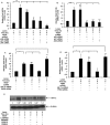
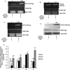
The potassium chloride cotransporters (KCC) family of proteins are widely expressed and are involved in the transepithelial movement of potassium and chloride ions and the regulation of cell volume. KCC activity is high in reticulocytes, and contributes to the dehydration of sickle red blood cells. Because plasma levels of both vascular endothelial growth factor (VEGF) and placental growth factor (PlGF) are elevated in sickle cell individuals, and VEGF has been shown to increase KCC expression in other cells, we hypothesized that VEGF and PlGF influence KCC expression in erythroid cells. Both VEGF and PlGF treatment of human erythroid K562 cells increased both mRNA and protein levels of KCC1, KCC3b, and KCC4. VEGF- and PlGF-mediated cellular signaling involved VEGF-R1 and downstream effectors, specifically, PI-3 kinase, p38 MAP kinase, mTOR, NADPH-oxidase, JNK kinase, and HIF-1α. VEGF and PlGF-mediated transcription of KCC3b and KCC4 involved hypoxia response element (HRE) motifs in their promoters, as demonstrated by promoter analysis, EMSA and ChiP. These results were corroborated in vivo by adenoviral-mediated overexpression of PlGF in normal mice, which led to increased expression of mKCC3 and mKCC4 in erythroid precursors. Our studies show that VEGF and PlGF regulate transcription of KCC3b and KCC4 in erythroid cells via activation of HIF-1α, independent of hypoxia. These studies provide novel therapeutic targets for regulation of cell volume in RBC precursors, and thus, amelioration of dehydration in RBCs in sickle cell disease.
Copyright © 2013 Wiley Periodicals, Inc.
Figures


Similar articles
-
Erythropoietin-mediated expression of placenta growth factor is regulated via activation of hypoxia-inducible factor-1α and post-transcriptionally by miR-214 in sickle cell disease.Biochem J. 2015 Jun 15;468(3):409-23. doi: 10.1042/BJ20141138. Epub 2015 Apr 16. Biochem J. 2015. PMID: 25876995 Free PMC article.
-
K-Cl cotransporter gene expression during human and murine erythroid differentiation.J Biol Chem. 2011 Sep 2;286(35):30492-30503. doi: 10.1074/jbc.M110.206516. Epub 2011 Jul 6. J Biol Chem. 2011. PMID: 21733850 Free PMC article.
-
Placenta growth factor-induced early growth response 1 (Egr-1) regulates hypoxia-inducible factor-1alpha (HIF-1alpha) in endothelial cells.J Biol Chem. 2010 Jul 2;285(27):20570-9. doi: 10.1074/jbc.M110.119495. Epub 2010 May 6. J Biol Chem. 2010. PMID: 20448047 Free PMC article.
-
Regulation of placental vascular endothelial growth factor (VEGF) and placenta growth factor (PIGF) and soluble Flt-1 by oxygen--a review.Placenta. 2000 Mar-Apr;21 Suppl A:S16-24. doi: 10.1053/plac.1999.0524. Placenta. 2000. PMID: 10831117 Review.
-
Structure and function of placental growth factor.Trends Cardiovasc Med. 2002 Aug;12(6):241-6. doi: 10.1016/s1050-1738(02)00168-8. Trends Cardiovasc Med. 2002. PMID: 12242046 Review.
Cited by
-
Capillary nano-immunoassays: advancing quantitative proteomics analysis, biomarker assessment, and molecular diagnostics.J Transl Med. 2015 Jun 6;13:182. doi: 10.1186/s12967-015-0537-6. J Transl Med. 2015. PMID: 26048678 Free PMC article. Review.
-
Erythropoietin-mediated expression of placenta growth factor is regulated via activation of hypoxia-inducible factor-1α and post-transcriptionally by miR-214 in sickle cell disease.Biochem J. 2015 Jun 15;468(3):409-23. doi: 10.1042/BJ20141138. Epub 2015 Apr 16. Biochem J. 2015. PMID: 25876995 Free PMC article.
-
A Novel Small Peptide H-KI20 Inhibits Retinal Neovascularization Through the JNK/ATF2 Signaling Pathway.Invest Ophthalmol Vis Sci. 2021 Jan 4;62(1):16. doi: 10.1167/iovs.62.1.16. Invest Ophthalmol Vis Sci. 2021. PMID: 33439229 Free PMC article.
-
K+-Cl- cotransporter 1 (KCC1): a housekeeping membrane protein that plays key supplemental roles in hematopoietic and cancer cells.J Hematol Oncol. 2019 Jul 11;12(1):74. doi: 10.1186/s13045-019-0766-x. J Hematol Oncol. 2019. PMID: 31296230 Free PMC article. Review.
-
The Zn2+-sensing receptor, ZnR/GPR39, upregulates colonocytic Cl- absorption, via basolateral KCC1, and reduces fluid loss.Biochim Biophys Acta Mol Basis Dis. 2017 Apr;1863(4):947-960. doi: 10.1016/j.bbadis.2017.01.009. Epub 2017 Jan 16. Biochim Biophys Acta Mol Basis Dis. 2017. PMID: 28093242 Free PMC article.
References
-
- Gamba G. Molecular physiology and pathophysiology of electroneutral cation-chloride cotransporters. Physiol Rev. 2005;85:423–493. - PubMed
-
- Lauf PK, Adragna NC. K–Cl cotransport: Properties and molecular mechanism. Cell Physiol Biochem. 2000;10:341–354. - PubMed
-
- Mount DB, Gamba G. Renal potassium–chloride cotransporters. Curr Opin Nephrol Hypertens. 2001;10:685–691. - PubMed
-
- Payne JA, Stevenson TJ, Donaldson LF. Cloning and expression of a neuronal-specific K–Cl cotransporter in rat brain. Soc Neurosci Abstr. 1996;22:732. - PubMed
-
- Hiki K, D'Andrea RJ, Furze J, et al. Cloning, characterization, and chromosomal location of a novel human K+–Cl− cotransporter. J Biol Chem. 1999;274:10661–10667. - PubMed
Publication types
MeSH terms
Substances
Grants and funding
LinkOut - more resources
Full Text Sources
Other Literature Sources
Research Materials
Miscellaneous
