. 2013 Dec;140(24):4970-81.
doi: 10.1242/dev.099192. Epub 2013 Nov 13.

N-glycosylation requirements in neuromuscular synaptogenesis

Affiliations

N-glycosylation requirements in neuromuscular synaptogenesis

William Parkinson et al. Development. 2013 Dec.

Abstract

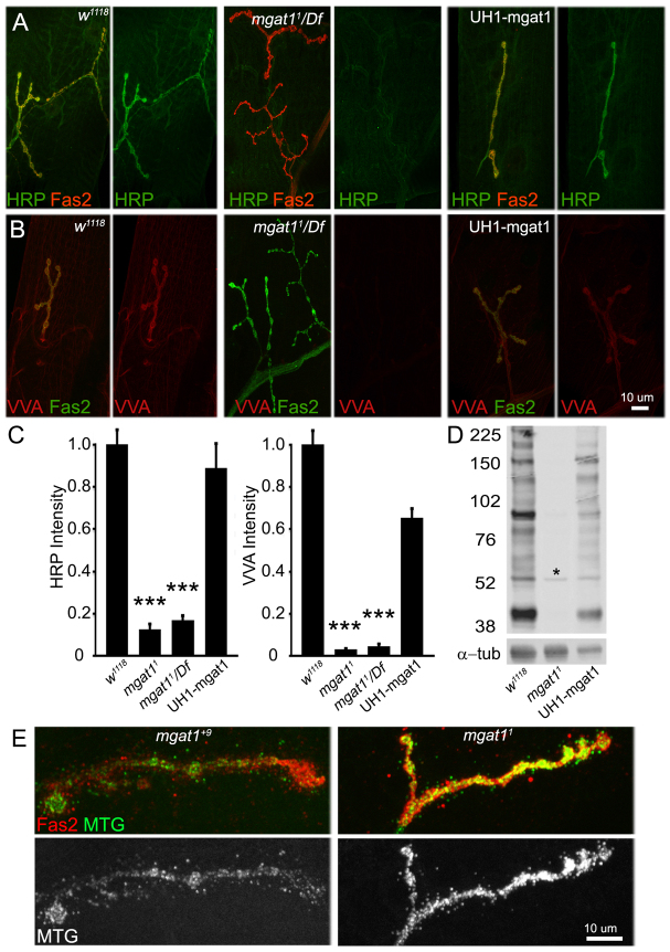
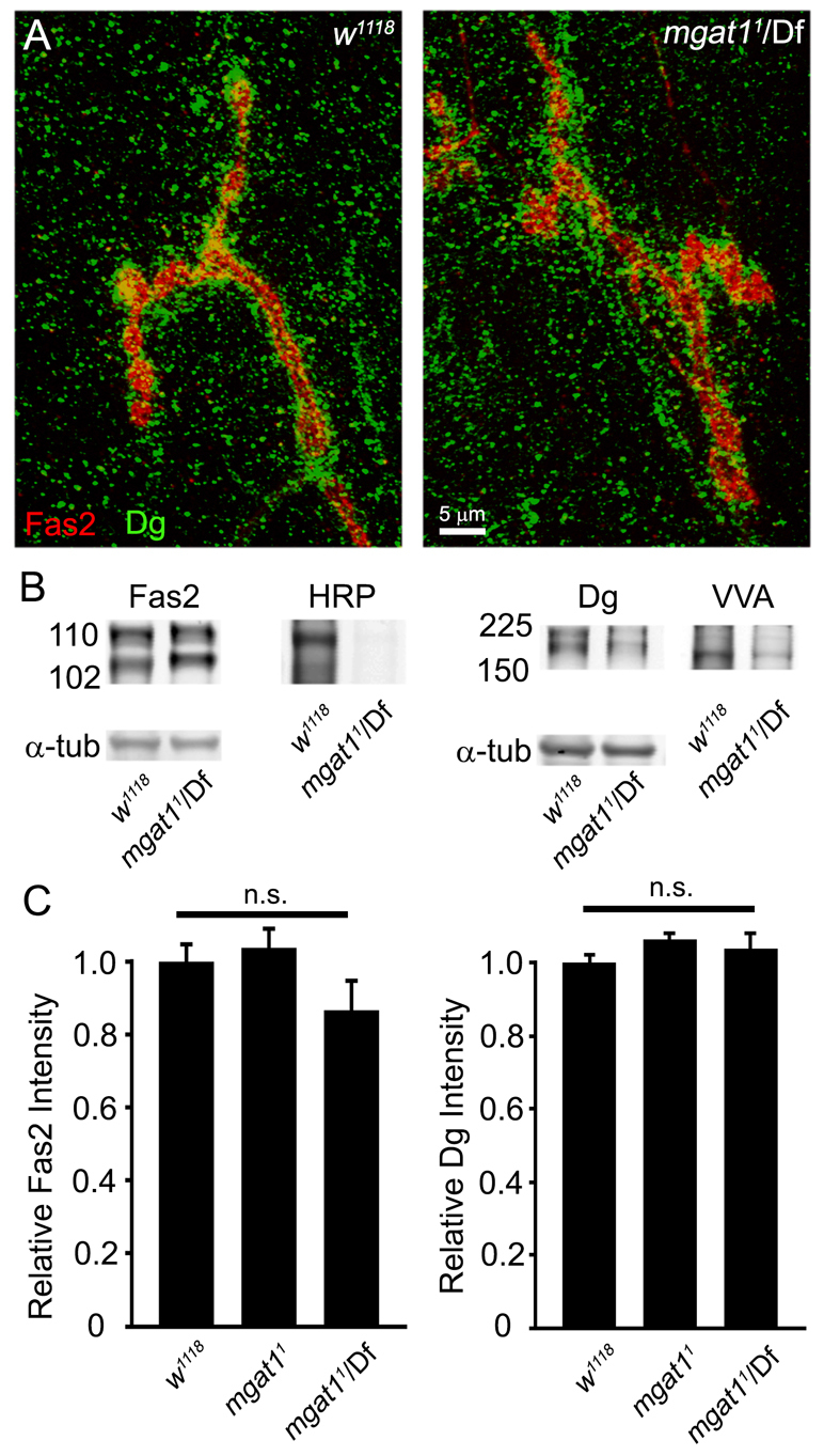
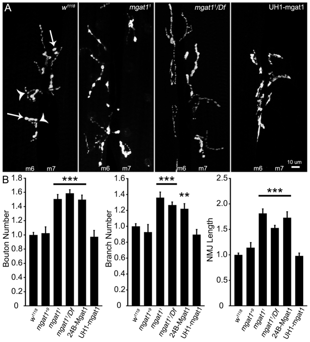
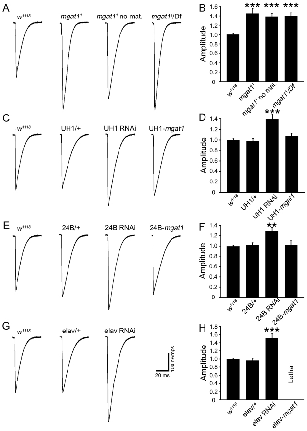
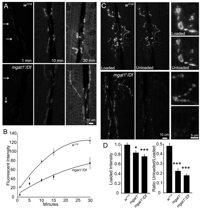
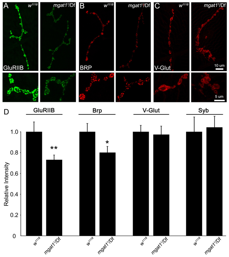
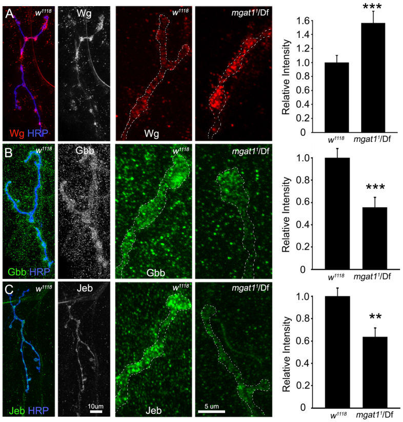
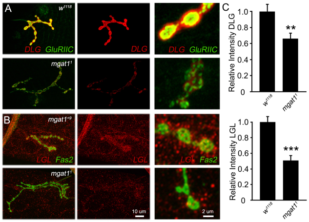
Neural development requires N-glycosylation regulation of intercellular signaling, but the requirements in synaptogenesis have not been well tested. All complex and hybrid N-glycosylation requires MGAT1 (UDP-GlcNAc:α-3-D-mannoside-β1,2-N-acetylglucosaminyl-transferase I) function, and Mgat1 nulls are the most compromised N-glycosylation condition that survive long enough to permit synaptogenesis studies. At the Drosophila neuromuscular junction (NMJ), Mgat1 mutants display selective loss of lectin-defined carbohydrates in the extracellular synaptomatrix, and an accompanying accumulation of the secreted endogenous Mind the gap (MTG) lectin, a key synaptogenesis regulator. Null Mgat1 mutants exhibit strongly overelaborated synaptic structural development, consistent with inhibitory roles for complex/hybrid N-glycans in morphological synaptogenesis, and strengthened functional synapse differentiation, consistent with synaptogenic MTG functions. Synapse molecular composition is surprisingly selectively altered, with decreases in presynaptic active zone Bruchpilot (BRP) and postsynaptic Glutamate receptor subtype B (GLURIIB), but no detectable change in a wide range of other synaptic components. Synaptogenesis is driven by bidirectional trans-synaptic signals that traverse the glycan-rich synaptomatrix, and Mgat1 mutation disrupts both anterograde and retrograde signals, consistent with MTG regulation of trans-synaptic signaling. Downstream of intercellular signaling, pre- and postsynaptic scaffolds are recruited to drive synaptogenesis, and Mgat1 mutants exhibit loss of both classic Discs large 1 (DLG1) and newly defined Lethal (2) giant larvae [L(2)GL] scaffolds. We conclude that MGAT1-dependent N-glycosylation shapes the synaptomatrix carbohydrate environment and endogenous lectin localization within this domain, to modulate retention of trans-synaptic signaling ligands driving synaptic scaffold recruitment during synaptogenesis.

Keywords: Active zone; Drosophila; Glutamate receptor; Neuromuscular junction; Synaptic scaffold; Synaptomatrix; Trans-synaptic signaling.


Figures

Fig. 1.
Fig. 1.
Loss of Mgat1 activity dramatically alters the Drosophila NMJ synaptomatrix. (A) Representative images of wandering third instar NMJs probed with anti-horseradish peroxidase (HRP, green) and anti-Fasciclin 2 (Fas2, red) in genetic control w1118, Mgat11/Df(2R)BSC430 and UH1-GAL4 driven UAS-Mgat1 in Mgat11 null background. HRP labeling is undetectable in the Mgat1 null, and fully restored by the genetic rescue. (B) Representative NMJ images of Vicia villosa (VVA, red) lectin labeling with Fas2 co-labeling (green) in the same genotypes. VVA labeling is undetectable in Mgat1 nulls, and fully restored by genetic rescue. (C) Quantification of HRP and VVA intensity normalized to w1118. ***P≤0.001 (ANOVA). Sample size is ≥10 NMJs from at least five animals of each genotype. (D) Representative anti-HRP western blot from w1118, Mgat1 null and UH1-Mgat1 rescue conditions. All HRP glycans are undetectable in Mgat1 null, and restored by Mgat1 rescue. The single band (asterisk) represents bleed-through from α-tubulin loading control. (E) Representative NMJ images of Mind the gap (MTG-GFP, green) lectin co-labeled with anti-Fas2 (red), and shown alone (MTG, white), in control (Mgat1+9 precise excision) and Mgat1 null. MTG is greatly increased in mutants (P≤0.0009; sample size: at least eight NMJs, at least four animals/genotype).
Fig. 2.
Fig. 2.
Fasciclin 2 and Dystroglycan are normally expressed in Mgat1 nulls. (A) Representative NMJ images of anti-Fasciclin 2 (Fas2, red) double-labeled with anti-Dystroglycan (DG, green) in w1118 control and Mgat11/Df mutant. (B) Representative western blots double-labeled for Fas2 and HRP (left) and DG and VVA lectin (right) in w1118 and Mgat11/Df. Alpha tubulin is the loading control. (C) Confocal fluorescence intensity quantification shows no significant (n.s.) change in Fas2 or DG at the NMJ in either homozygous Mgat11 or Mgat11/Df conditions compared with w1118. ANOVA statistical analyses were carried out on a sample size of n≥16 NMJs for each genotype.
Fig. 3.
Fig. 3.
Drosophila NMJ is structurally overgrown in Mgat1 null mutants. NMJ length, axon branching and synaptic bouton number are increased in Mgat1 nulls. (A) Representative images of wandering third instar 6/7 NMJ in genetic control (w1118), Mgat11 null, Mgat11/Df(2)BSC430 and UH1-Gal4 driven UAS-Mgat1 in null background. Representative axon branch points (arrowheads) and boutons (arrows) are illustrated. (B) Quantification of bouton number, branch number and NMJ length normalized to w1118 for the Mgat1+9 precise excision control, Mgat11 homozygous null, Mgat11/Df and UAS-Mgat1 driven in muscle (24B-Gal4) or ubiquitously (UH1-Gal4) in the Mgat11/Df background. All three structural parameters are increased in Mgat1 nulls and fully rescued by ubiquitous UAS-Mgat1 expression, but not muscle-targeted expression. **P≤0.01 and ***P≤0.001 (ANOVA) compared with w1118. Sample size is at least eight NMJs from at least four animals from each of the six genotypes shown.
Fig. 4.
Fig. 4.
Strengthened synaptic functional differentiation in Mgat1 null mutants. Neurotransmission measured using TEVC of stimulation-evoked EJCs at the wandering third instar muscle 6 NMJ. (A) Representative EJC traces recorded in 1.0 mM Ca2+ from w1118, Mgat11 null, Mgat11 without maternal contribution and Mgat11/Df. (B) EJC quantification for all four genotypes normalized to genetic control. Sample size is at least tn animals per genotype. (C) Traces from w1118, UH1/+ control, UH1-GAL4 driven UAS-Mgat1-RNAi and UH1-GAL4 driven UAS-Mgat1 in Mgat11/Df background. Synaptic transmission is elevated by ubiquitous RNAi knockdown and fully restored with reintroduction of ubiqutousUAS-Mgat1. (D) EJC quantification for all four genotypes normalized to w1118. Sample size is ≥14 animals per genotype. (E) Traces from w1118, 24B-GAL4/+ control, 24B-GAL4 driven UAS-Mgat1-RNAi and 24B-GAL4 driven UAS-Mgat1 in null background. Transmission elevated by postsynaptic RNAi and rescued with postsynaptic UAS-Mgat1. (F) EJC quantification for four genotypes normalized to w1118. Sample size is ≥11 animals per genotype. (G) Traces from w1118, elav-GAL4/+ control and elav-GAL4 driven UAS-Mgat1-RNAi. Transmission is elevated with neuronal RNAi knockdown. (H) EJC quantification normalized to w1118. Sample size is ≥11 animals per genotype. **P≤0.01 and ***P≤0.001 (ANOVA).
Fig. 5.
Fig. 5.
Altered NMJ synaptic vesicle cycling in the Mgat1 null mutants. Assays of synaptic vesicle (SV) loading and cycling rates with FM1-43 dye imaging at the wandering third instar 6/7 NMJ. (A) Representative images of FM1-43 loading during endogenous activity at 1, 10 and 30 minutes. Top panels show w1118 control and bottom panels show Mgat11/Df. Arrows indicate boutons faintly loaded at 1 minute. (B) Fluorescence intensity plotted at 1, 5, 10, 15 and 30 minutes of endogenous activity; at least nine animals per genotype used for each time point. (C) Representative NMJ images after FM1-43 loading with 90mM K+ for 5 minutes and unloading for 2 minutes. Top panels show w1118 and bottom panels show Mgat11/Df, with increased SV turnover indicated by reduced intensity after unloading. Right panels show high magnification images of loaded/unloaded boutons. (D) Quantification of loading and ratio of unloaded/loaded fluorescence intensities for w1118, Mgat11 homozygous null and Mgat11/Df. *P≤0.05, **P≤0.01 and ***P≤0.001 (ANOVA) compared with genetic control w1118. Sample size is ≥18 NMJs for each genotype.
Fig. 6.
Fig. 6.
Pre- and postsynaptic component recruitment reduced at Mgat1 NMJs. Null Mgat1 mutants show decreased postsynaptic glutamate receptor type IIB (GLURIIB) and presynaptic active zone Bruchpilot (BRP), but are unchanged for multiple other synaptic components. (A-C) Representative NMJ images for GLURIIB (A, green), BRP (B, red) and the vesicular glutamate transporter (VGLUT; C, red) in genetic control w1118 and Mgat11/Df mutants. High-magnification images of synaptic boutons shown below. (D) Quantification of fluorescence intensities for GLURIIB (n≥28), BRP (n≥38), VGLUT (n≥12) and Synaptobrevin (SYB; n≥16) normalized to w1118. VGLUT and SYB exhibit no change, indicating similar synaptic vesicle related density in control and mutant. *P≤0.05, **P≤0.01 and ***P≤0.001 (Student’s t-test) for each pairwise comparison.
Fig. 7.
Fig. 7.
Multiple trans-synaptic signaling pathways altered in Mgat1 null mutant. Assays of three well-characterized NMJ trans-synaptic signals: Wingless (WG), Glass bottom boat (GBB) and Jelly belly (JEB). (A) Representative NMJ images of WG (red) double-labeled with HRP (blue), and shown alone (WG, white). High-magnification bouton comparison of genetic control (w1118) and Mgat11/Df. Right: Quantification of relative fluorescence intensity reveals increased WG in mutant. (B) Representative images of GBB (green) double-labeled with HRP (blue), and shown alone (GBB, white). High-magnification bouton comparison of w1118 and Mgat11/Df. Right: Quantification shows decreased GBB in mutant. (C) Representative NMJ images of JEB (green) double-labeled with HRP (blue), and shown alone (JEB, white). High-magnification bouton comparison of w1118 and Mgat11/Df. Right: Quantification shows decreased JEB in mutant. Fluorescence intensities measured within the NMJ domain (white dotted line), normalized to genetic control w1118. **P≤0.01 and ***P≤0.001 (Student’s t-test) for pairwise comparisons. The sample size is n≥18 NMJs for each label and each genotype.
Fig. 8.
Fig. 8.
Synaptic scaffolds DLG1 and L(2)GL reduced in the Mgat1 null mutants. Imaging of DLG1 and L(2)GL synaptic scaffolds at the wandering third instar NMJ. (A) Representative images of DLG1 (red) co-labeled with glutamate receptor type IIC (GLURIIC, green), with higher magnification boutons in side panels. (B) Representative images of L(2)GL (red) co-labeled with Fas2 (green), with high-magnification boutons in side panels. (C) Quantification of DLG1 fluorescence intensity (top: Mgat11, n=15; w1118, n=22) and L(2)GL fluorescence intensity (bottom: Mgat11, n=12; w1118, n=12). **P≤0.01 and ***P≤0.001 (Student’s t-test) for pairwise comparisons.

References

    1. Abramoff M. D., Magalhaes P. J., Ram S. J. (2004). Image processing with ImageJ. Biophotonics International 11, 36–42
    1. Ataman B., Ashley J., Gorczyca D., Gorczyca M., Mathew D., Wichmann C., Sigrist S. J., Budnik V. (2006). Nuclear trafficking of Drosophila Frizzled-2 during synapse development requires the PDZ protein dGRIP. Proc. Natl. Acad. Sci. USA 103, 7841–7846 - PMC - PubMed
    1. Ataman B., Ashley J., Gorczyca M., Ramachandran P., Fouquet W., Sigrist S. J., Budnik V. (2008). Rapid activity-dependent modifications in synaptic structure and function require bidirectional Wnt signaling. Neuron 57, 705–718 - PMC - PubMed
    1. Beck E. S., Gasque G., Imlach W. L., Jiao W., Jiwon Choi B., Wu P. S., Kraushar M. L., McCabe B. D. (2012). Regulation of Fasciclin II and synaptic terminal development by the splicing factor beag. J. Neurosci. 32, 7058–7073 - PMC - PubMed
    1. Beumer K. J., Rohrbough J., Prokop A., Broadie K. (1999). A role for PS integrins in morphological growth and synaptic function at the postembryonic neuromuscular junction of Drosophila. Development 126, 5833–5846 - PubMed

Publication types

MeSH terms

LinkOut - more resources