

Changes in brain function occur years before the onset of cognitive impairment
- PMID: 24227712
- PMCID: PMC3828456
- DOI: 10.1523/JNEUROSCI.1402-13.2013
Changes in brain function occur years before the onset of cognitive impairment
Abstract
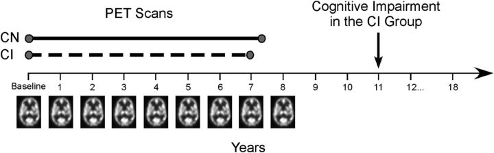
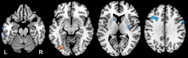
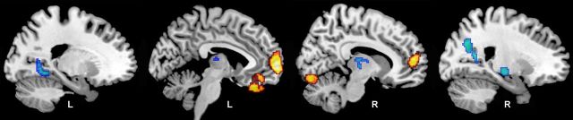
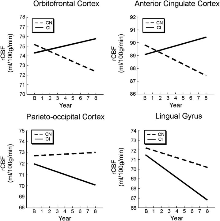
To develop targeted intervention strategies for the treatment of Alzheimer's disease, we first need to identify early markers of brain changes that occur before the onset of cognitive impairment. Here, we examine changes in resting-state brain function in humans from the Baltimore Longitudinal Study of Aging. We compared longitudinal changes in regional cerebral blood flow (rCBF), assessed by (15)O-water PET, over a mean 7 year period between participants who eventually developed cognitive impairment (n = 22) and those who remained cognitively normal (n = 99). Annual PET assessments began an average of 11 years before the onset of cognitive impairment in the subsequently impaired group, so all participants were cognitively normal during the scanning interval. A voxel-based mixed model analysis was used to compare groups with and without subsequent impairment. Participants with subsequent impairment showed significantly greater longitudinal rCBF increases in orbitofrontal, medial frontal, and anterior cingulate regions, and greater longitudinal decreases in parietal, temporal, and thalamic regions compared with those who maintained cognitive health. These changes were linear in nature and were not influenced by longitudinal changes in regional tissue volume. Although all participants were cognitively normal during the scanning interval, most of the accelerated rCBF changes seen in the subsequently impaired group occurred within regions thought to be critical for the maintenance of cognitive function. These changes also occurred within regions that show early accumulation of pathology in Alzheimer's disease, suggesting that there may be a connection between early pathologic change and early changes in brain function.
Figures
References
-
- Andrews-Zwilling Y, Bien-Ly N, Xu Q, Li G, Bernardo A, Yoon SY, Zwilling D, Yan TX, Chen L, Huang Y. Apolipoprotein E4 causes age- and Tau-dependent impairment of GABAergic interneurons, leading to learning and memory deficits in mice. J Neurosci. 2010;30:13707–13717. doi: 10.1523/JNEUROSCI.4040-10.2010. - DOI - PMC - PubMed
-
- Axmacher N, Schmitz DP, Wagner T, Elger CE, Fell J. Interactions between medial temporal lobe, prefrontal cortex, and inferior temporal regions during visual working memory: a combined intracranial EEG and functional magnetic resonance imaging study. J Neurosci. 2008;28:7304–7312. doi: 10.1523/JNEUROSCI.1778-08.2008. - DOI - PMC - PubMed
-
- Bateman RJ, Xiong C, Benzinger TL, Fagan AM, Goate A, Fox NC, Marcus DS, Cairns NJ, Xie X, Blazey TM, Holtzman DM, Santacruz A, Buckles V, Oliver A, Moulder K, Aisen PS, Ghetti B, Klunk WE, McDade E, Martins RN, et al. Clinical and biomarker changes in dominantly inherited Alzheimer's disease. N Engl J Med. 2012;367:795–804. doi: 10.1056/NEJMoa1202753. - DOI - PMC - PubMed
Publication types
MeSH terms
Grants and funding
LinkOut - more resources
Full Text Sources
Other Literature Sources
Medical