. 2013 Dec 15;191(12):6250-60.
doi: 10.4049/jimmunol.1301728. Epub 2013 Nov 13.

MicroRNAs transfer from human macrophages to hepato-carcinoma cells and inhibit proliferation

Affiliations

MicroRNAs transfer from human macrophages to hepato-carcinoma cells and inhibit proliferation

Anne Aucher et al. J Immunol. .

Abstract

Recent research has indicated a new mode of intercellular communication facilitated by the movement of RNA between cells. There is evidence that RNA can transfer between cells in a multitude of ways, including in complex with proteins or lipids or in vesicles, including apoptotic bodies and exosomes. However, there remains little understanding of the function of nucleic acid transfer between human cells. In this article, we report that human macrophages transfer microRNAs (miRNAs) to hepato-carcinoma cells (HCCs) in a manner that required intercellular contact and involved gap junctions. Two specific miRNAs transferred efficiently between these cells--miR-142 and miR-223--and both were endogenously expressed in macrophages and not in HCCs. Transfer of these miRNAs influenced posttranscriptional regulation of proteins in HCCs, including decreased expression of reporter proteins and endogenously expressed stathmin-1 and insulin-like growth factor-1 receptor. Importantly, transfer of miRNAs from macrophages functionally inhibited proliferation of these cancerous cells. Thus, these data led us to propose that intercellular transfer of miRNA from immune cells could serve as a new defense against unwanted cell proliferation or tumor growth.


Figures
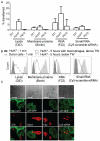
Figure 1
Figure 1. Macrophages transfer cell components, including RNA molecules, to hepato-carcinoma cells
(A) Macrophages were stained with the fluorescent lipid DiD or biotin or RNA dye F22, or were transfected with Cy5-scramble-siRNA, and co-cultured with 221, HuH7 or P815 transfected to express GPI-GFP for 1 min. or 5 h. Graph shows the percentage of fluorescence initially present in macrophages that had transferred to recipient cells as determined by flow cytometry. Error bars are SEM. n = 3. (B) HuH7 were cultured above or below a transwell membrane (TW), with macrophages stained as described in (A), added only to the compartment above the TW. After 1 min. or 5 h of co-culture, cells were analysed by flow cytometry. Light grey histogram shows the level of fluorescence in the donor cells at the beginning of the co-culture. Data are representative of 4 donors. (C) Donor macrophages were stained as described in (A) (red) and co-cultured with HuH7 (marked by GFP, green). After 5 h of co-culture, cells were analysed by confocal microscopy. Scale bars: 10 μm. n > 44 cells for each condition, from 6 independent experiments.
Figure 2
Figure 2. Specific miRNAs are transferred from macrophages to HCCs
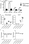
Relative expression of miR-142, -223, -425 and -122 was assessed by quantitative Real-Time PCR (qRT-PCR) in macrophages and HuH7 alone (A). Fold change in the level of expression of miR-142, -223, -425 and -122 was assessed by quantitative Real-Time PCR (qRT-PCR) in isolated macrophages and HuH7 that had been co-cultured together for 1 min. or 5 h - macrophages are represented in (B) and HuH7 in (C). Data points represent individual experiments in (A); (B) n = 3; (c) n > 5. (D) Relative expression of each miRNA in HuH7, after 1 min. or 5 h of co-culture with macrophages was individually plotted. Connected data points represent individual experiments, each using cells from a different donor. Expression of miR-142, -223, -425 and -122 in primary human B and T cells alone (E) or in HuH7 co-cultured with T or B cells for 1 min. or 5 h (F). Data points represent individual donors in (E); (F) B cells: n = 3, T cells: n = 4. Error bars are SEM. Significance was determined by a non-parametric Mann-Whitney U test.
Figure 3
Figure 3. miR-142 and -223 are not newly synthesised in HuH7 following co-culture with macrophages
(A) Fold change in the level of miR-142, -223, -425 and -122 present in HuH7 cells, assessed by qRT-PCR, following co-culture with macrophages that were either alive or fixed in paraformaldehyde (PFA). n = 4. (B) Relative expression of precursors of miR-142, -223, -425 and -122 in macrophages, and HuH7 analysed after 5 h of co-culture. Data points represent individual experiments. (C) Fold change of pri-miRNAs in macrophages and HuH7 cells following their co-culture. n > 3. N.D. - Not Detected. Error bars are SEM. Significance was determined by a non-parametric Mann-Whitney U test.
Figure 4
Figure 4. miRNAs are transferred via a direct contact between macrophages and HCCs
(A) Percent of cells containing apoptotic bodies, as stained by Draq5 and detected by confocal microscopy, for HuH7 alone or co-cultured with macrophages or co-cultured with apoptotic bodies isolated from macrophages treated with cycloheximide (CHX). n > 707 cells for each condition, from 6 independent experiments. (B) Fold change in level of miR-142 and -223, assessed by qRT-PCR, in HuH7 co-cultured for 5 h with isolated apoptotic bodies derived from macrophages treated with CHX. n = 4. (C) Fold change in the level of miR-142 and -223 in HuH7, as determined by qRT-PCR, following co-culture for 5 h with macrophages, macrophages in presence of 10 μg/ml brefeldin A (Bref A), or with exosomes that were isolated from cultures of macrophages or isolated from macrophages co-cultured with HuH7. n = 4. (D) Fold change of miR-142 and -223 in HuH7 co-cultured for 5 h with macrophages, supernatant form macrophages or supernatant from macrophages previously co-cultured with HuH7, in the presence or absence of an antibody blocking HDL receptors or 5 μM manumycin A. n > 4. (E) Fold change of miR-142 and -223 in HuH7 co-cultured for 5 h above or below a TW, with macrophages added only to compartment above the TW. n > 4. (F) Fold change of miR-142 and -223 in HuH7 co-cultured for 5 h with macrophages after being left alone or treated with 1 μM latrunculin A or 5 μM nocodazole. n = 4. (G) As in (F) except that cells were either left alone or pre-treated and then co-cultured with 75 μM 18-alpha-GA or 1 mM 2-octanol or 100 μM oleamide. n = 4. Error bars are SEM. Significance was determined by a non-parametric Mann-Whitney U test.
Figure 5
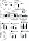
Figure 5. Intercellular transfer of miRNAs is functionally important
(A) Fold change of STMN1 and IGF1-R mRNA was assessed by qRT-PCR in HuH7 cells alone or co-cultured with macrophages for 5 or 24 h. n = 6. (B) As in (A), except that HuH7 cells were stably transfected to express anti-miR-223 sponges, or control scramble sponges, and analysed for the expression of STMN1 mRNA. n = 5. (C) HuH7 cells, transfected to express a plasmid encoding the wild-type, a control truncated or mutated 3′UTR of STMN1 fused to the firefly luciferase, along with another plasmid encoding the Renilla luciferase, were co-cultured alone or with macrophages that were either alive or fixed in PFA. Luciferase activity was assessed after 5 or 24 h. n = 6. (D) As in (C), but HuH7 were stably transfected to express anti-miR-223 sponges or control scramble sponges. n = 6. (E) As in (C), but macrophages were transfected to express anti-miR-223 antagomiRs or control scramble antagomiRs. n = 4. (F) Proliferation of HuH7 or HepG2 co-cultured with live or fixed macrophages was measured by incorporation of [3H]-thymidine over 4 days. Graph shows proliferation of HCCs in co-cultures relative to HCCs cultured alone. n > 7. (G) As in (F), except that where indicated, macrophages and HuH7 were pre-treated for 24 h with 75 μM 18-alpha-GA. (H) As in (F), except that HCCs were stably transfected to express anti-miR-223 sponges or control scramble sponges. n = 3. (I) As in (F), but macrophages were transfected with anti-miR-223 antagomiRs or control scramble antagomiRs. n = 4. Error bars are SEM. Significance was determined by a non-parametric Mann-Whitney U test.

References

    1. Fishman M. Antibody formation in vitro. J. Exp. Med. 1961;114:837–856. - PMC - PubMed
    1. Chitwood DH, Timmermans MCP. Small RNAs are on the move. Nature. 2010;467:415–419. - PubMed
    1. Vickers KC, Palmisano BT, Shoucri BM, Shamburek RD, Remaley AT. MicroRNAs are transported in plasma and delivered to recipient cells by high-density lipoproteins. Nature Cell Biology. 2011;13:423–433. - PMC - PubMed
    1. Zernecke A, Bidzhekov K, Noels H, Shagdarsuren E, Gan L, Denecke B, Hristov M, Köppel T, Jahantigh MN, Lutgens E, Wang S, Olson EN, Schober A, Weber C. Delivery of microRNA-126 by apoptotic bodies induces CXCL12-dependent vascular protection. Science Signaling. 2009;2:ra81. - PubMed
    1. Valadi H, Ekström K, Bossios A, Sjöstrand M, Lee JJ, Lötvall JO. Exosome-mediated transfer of mRNAs and microRNAs is a novel mechanism of genetic exchange between cells. Nature Cell Biology. 2007;9:654–659. - PubMed

Publication types

MeSH terms