

Epithelial NF-κB orchestrates house dust mite-induced airway inflammation, hyperresponsiveness, and fibrotic remodeling
- PMID: 24227776
- PMCID: PMC3858534
- DOI: 10.4049/jimmunol.1301329
Epithelial NF-κB orchestrates house dust mite-induced airway inflammation, hyperresponsiveness, and fibrotic remodeling
Abstract
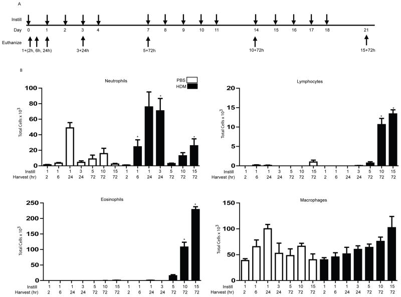
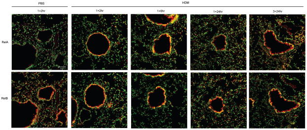
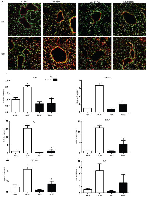
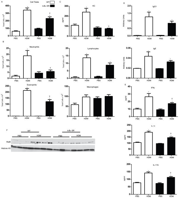
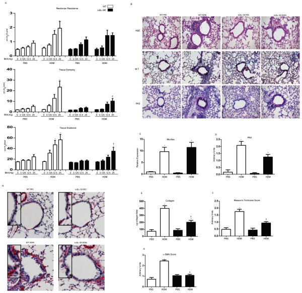
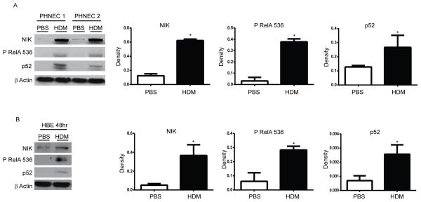
NF-κB activation within the epithelium has been implicated in the pathogenesis of asthma, yet the exact role of epithelial NF-κB in allergen-induced inflammation and airway remodeling remains unclear. In the current study, we used an intranasal house dust mite (HDM) extract exposure regimen time course in BALB/c mice to evaluate inflammation, NF-κB activation, airway hyperresponsiveness (AHR), and airway remodeling. We used CC10-IκBαSR transgenic mice to evaluate the functional importance of epithelial NF-κB in response to HDM. After a single exposure of HDM, mRNA expression of proinflammatory mediators was significantly elevated in lung tissue of wild-type (WT) mice, in association with increases in nuclear RelA and RelB, components of the classical and alternative NF-κB pathway, respectively, in the bronchiolar epithelium. In contrast, CC10-IκBαSR mice displayed marked decreases in nuclear RelA and RelB and mRNA expression of proinflammatory mediators compared with WT mice. After 15 challenges with HDM, WT mice exhibited increases in inflammation, AHR, mucus metaplasia, and peribronchiolar fibrosis. CC10-IκBαSR transgenic mice displayed marked decreases in neutrophilic infiltration, tissue damping, and elastance parameters, in association will less peribronchiolar fibrosis and decreases in nuclear RelB in lung tissue. However, central airway resistance and mucus metaplasia remained elevated in CC10-IκBαSR transgenic mice, in association with the continued presence of lymphocytes, and partial decreases in eosinophils and IL-13. The current study demonstrates that following airway exposure with an asthma-relevant allergen, activation of classical and alternative NF-κB pathways occurs within the airway epithelium and may coordinately contribute to allergic inflammation, AHR, and fibrotic airway remodeling.
Figures
Similar articles
-
Therapeutic targets for inflammation-mediated airway remodeling in chronic lung disease.Expert Rev Respir Med. 2018 Nov;12(11):931-939. doi: 10.1080/17476348.2018.1526677. Epub 2018 Oct 3. Expert Rev Respir Med. 2018. PMID: 30241450 Free PMC article. Review.
-
NF-κB/RelA signaling in secretoglobin progenitors mediates plasticity and MMP-induced barrier disruption in house dust mite-induced allergic asthma.Am J Physiol Lung Cell Mol Physiol. 2024 Jul 1;327(1):L86-L101. doi: 10.1152/ajplung.00066.2024. Epub 2024 May 7. Am J Physiol Lung Cell Mol Physiol. 2024. PMID: 38713619 Free PMC article.
-
Distinct functions of airway epithelial nuclear factor-kappaB activity regulate nitrogen dioxide-induced acute lung injury.Am J Respir Cell Mol Biol. 2010 Oct;43(4):443-51. doi: 10.1165/rcmb.2008-0416OC. Epub 2009 Nov 9. Am J Respir Cell Mol Biol. 2010. PMID: 19901348 Free PMC article.
-
A prominent role for airway epithelial NF-kappa B activation in lipopolysaccharide-induced airway inflammation.J Immunol. 2003 Jun 15;170(12):6257-65. doi: 10.4049/jimmunol.170.12.6257. J Immunol. 2003. PMID: 12794158
-
Cytokines and growth factors in airway remodeling in asthma.Curr Opin Immunol. 2007 Dec;19(6):676-80. doi: 10.1016/j.coi.2007.07.017. Epub 2007 Aug 27. Curr Opin Immunol. 2007. PMID: 17720466 Review.
Cited by
-
NF-kappaB Signaling in Chronic Inflammatory Airway Disease.Biomolecules. 2015 Jun 26;5(3):1266-83. doi: 10.3390/biom5031266. Biomolecules. 2015. PMID: 26131974 Free PMC article. Review.
-
The Kiwifruit Allergen Act d 1 Activates NF-κB Signaling and Affects mRNA Expression of TJ Proteins and Innate Pro-Allergenic Cytokines.Biomolecules. 2019 Dec 2;9(12):816. doi: 10.3390/biom9120816. Biomolecules. 2019. PMID: 31810340 Free PMC article.
-
Therapeutic targets for inflammation-mediated airway remodeling in chronic lung disease.Expert Rev Respir Med. 2018 Nov;12(11):931-939. doi: 10.1080/17476348.2018.1526677. Epub 2018 Oct 3. Expert Rev Respir Med. 2018. PMID: 30241450 Free PMC article. Review.
-
Aspergillus fumigatus Drives Tissue Damage via Iterative Assaults upon Mucosal Integrity and Immune Homeostasis.Infect Immun. 2023 Feb 16;91(2):e0033322. doi: 10.1128/iai.00333-22. Epub 2023 Jan 10. Infect Immun. 2023. PMID: 36625602 Free PMC article.
-
Chinese herbal component, Praeruptorin E, enhances anti-asthma efficacy and prevents toxicity of aminophylline by targeting the NF-κB/PXR/CYP3A4 pathway.Ann Transl Med. 2022 Feb;10(4):225. doi: 10.21037/atm-22-386. Ann Transl Med. 2022. PMID: 35280431 Free PMC article.
References
-
- Hayden MS, Ghosh S. Signaling to NF-kappaB. Genes Dev. 2004;18:2195–2224. - PubMed
-
- Scheidereit C. IkappaB kinase complexes: gateways to NF-kappaB activation and transcription. Oncogene. 2006;25:6685–6705. - PubMed
-
- Senftleben U, Cao Y, Xiao G, Greten FR, Krahn G, Bonizzi G, Chen Y, Hu Y, Fong A, Sun SC, Karin M. Activation by IKKalpha of a second, evolutionary conserved, NF-kappa B signaling pathway. Science. 2001;293:1495–1499. - PubMed
-
- Oeckinghaus A, Hayden MS, Ghosh S. Crosstalk in NF-kappaB signaling pathways. Nat Immunol. 2011;12:695–708. - PubMed
Publication types
MeSH terms
Substances
Grants and funding
LinkOut - more resources
Full Text Sources
Other Literature Sources
Molecular Biology Databases
Miscellaneous
