

Small incision lenticule extraction (SMILE) and femtosecond laser LASIK: comparison of corneal wound healing and inflammation
- PMID: 24227802
- PMCID: PMC3913294
- DOI: 10.1136/bjophthalmol-2013-303415
Small incision lenticule extraction (SMILE) and femtosecond laser LASIK: comparison of corneal wound healing and inflammation
Abstract
Aim: To evaluate and compare early corneal wound healing and inflammatory responses after small incision lenticule extraction (SMILE) versus femtosecond laser laser in situ keratomileusis (LASIK).
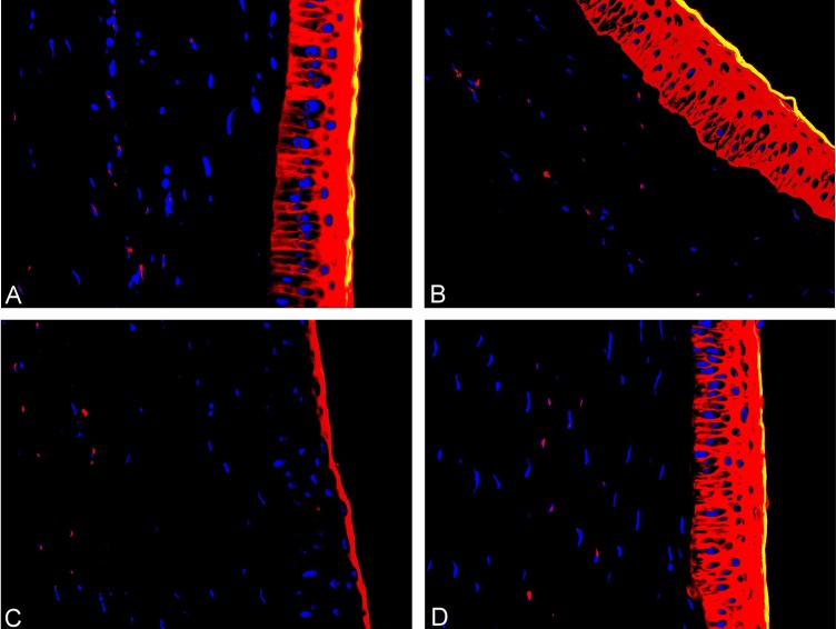
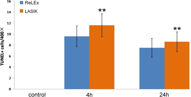
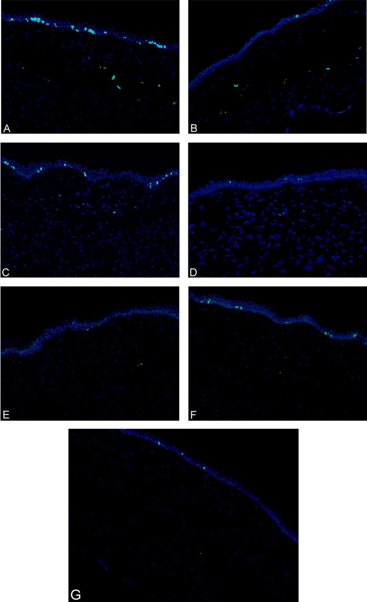
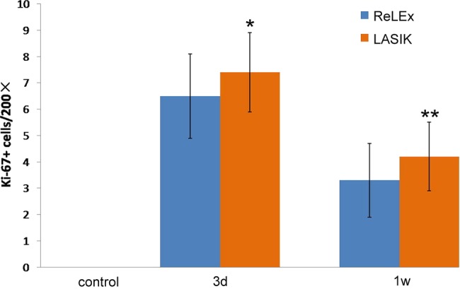
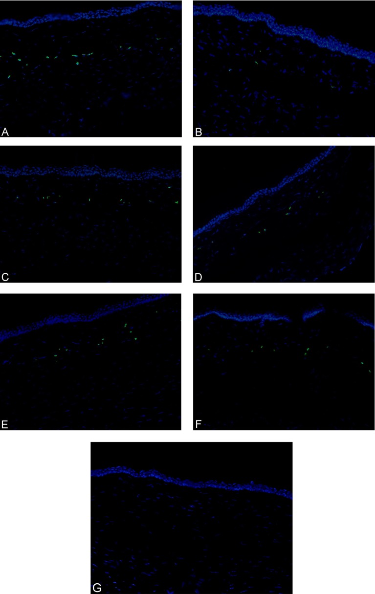
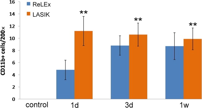
Methods: Thirty-six eyes of 36 rabbits underwent SMILE, while another 36 eyes of 36 rabbits were treated with femtosecond laser LASIK. All the eyes were subjected to the same refractive correction of -6.00 DS/-1.00 DC. Twelve eyes that had no surgery were included for control. After euthanisation, corneal tissue sections were evaluated with terminal deoxyribonucleotidyl transferase-mediated dUTP-digoxigenin nick-end labelling (TUNEL) assay to detect apoptosis at postoperative 4 and 24 h, immunocytochemistry for Ki67 to detect keratocyte proliferation at postoperative day 3, week 1 and month 1, and immunocytochemistry for CD11b to detect inflammation at postoperative day 1, day 3 and week 1, respectively.
Results: No adverse effects were noted after SMILE or LASIK. Corneal healing postoperatively was uneventful in all cases. There were significantly fewer TUNEL-positive corneal stromal cells after the SMILE procedure at 4 and 24 h postoperatively (p<0.01) compared with the LASIK procedure. In addition, immunocytochemistry showed significantly fewer Ki67-positive cells in the SMILE group than those in the femtosecond laser LASIK group at day 3 and week 1 postoperatively (p<0.05), but there was little expression of Ki67 at month 1 postoperatively in both groups. The CD11b-positive cells were significantly fewer in the SMILE group at day 1, day 3 and week 1 postoperatively (p<0.01).
Conclusions: SMILE induces less keratocyte apoptosis, proliferation and inflammation compared with femtosecond laser LASIK.
Keywords: Cornea; Inflammation; Wound Healing.
Figures
Similar articles
-
Wound Healing, Inflammation, and Corneal Ultrastructure After SMILE and Femtosecond Laser-Assisted LASIK: A Human Ex Vivo Study.J Refract Surg. 2018 Jun 1;34(6):393-399. doi: 10.3928/1081597X-20180425-02. J Refract Surg. 2018. PMID: 29889292
-
Comparison of Corneal Biological Healing After Femtosecond LASIK and Small Incision Lenticule Extraction Procedure.Curr Eye Res. 2016 Sep;41(9):1202-8. doi: 10.3109/02713683.2015.1107590. Epub 2016 Feb 1. Curr Eye Res. 2016. PMID: 26833247 Clinical Trial.
-
[The histopathological observation of rabbit corneas after small incision lenticule extraction].Zhonghua Yan Ke Za Zhi. 2016 Jul;52(7):507-13. doi: 10.3760/cma.j.issn.0412-4081.2016.07.008. Zhonghua Yan Ke Za Zhi. 2016. PMID: 27531111 Clinical Trial. Chinese.
-
Femtosecond laser refractive surgery: small-incision lenticule extraction vs. femtosecond laser-assisted LASIK.Curr Opin Ophthalmol. 2015 Jul;26(4):260-4. doi: 10.1097/ICU.0000000000000158. Curr Opin Ophthalmol. 2015. PMID: 26058022 Review.
-
Dry Eye After Small Incision Lenticule Extraction and Femtosecond Laser-Assisted LASIK: Meta-Analysis.Cornea. 2017 Jan;36(1):85-91. doi: 10.1097/ICO.0000000000000999. Cornea. 2017. PMID: 27560032 Review.
Cited by
-
In vivo confocal microscopy of sub-basal corneal nerves and corneal densitometry after three kinds of refractive procedures for high myopia.Int Ophthalmol. 2023 Mar;43(3):925-935. doi: 10.1007/s10792-022-02494-0. Epub 2022 Sep 25. Int Ophthalmol. 2023. PMID: 36153757
-
Biology of keratorefractive surgery- PRK, PTK, LASIK, SMILE, inlays and other refractive procedures.Exp Eye Res. 2020 Sep;198:108136. doi: 10.1016/j.exer.2020.108136. Epub 2020 Jul 10. Exp Eye Res. 2020. PMID: 32653492 Free PMC article. Review.
-
Smile--the next generation of laser vision correction.Rom J Ophthalmol. 2016 Jan-Mar;60(1):6-8. Rom J Ophthalmol. 2016. PMID: 27220224 Free PMC article.
-
Clinical Outcomes of Small Incision Lenticule Extraction in Myopia: Study of Vector Parameters and Corneal Aberrations.Korean J Ophthalmol. 2020 Feb;34(1):76-84. doi: 10.3341/kjo.2019.0109. Korean J Ophthalmol. 2020. PMID: 32037752 Free PMC article.
-
The Effect of High Intensity Focused Ultrasound Keratoplasty on Rabbit Anterior Segment.J Ophthalmol. 2017;2017:6067890. doi: 10.1155/2017/6067890. Epub 2017 Feb 9. J Ophthalmol. 2017. PMID: 28280636 Free PMC article.
References
-
- Soong HK, Malta JB. Femtosecond lasers in ophthalmology. Am J Ophthalmol 2009;147:189–97 - PubMed
-
- Kim P, Sutton GL, Rootman DS. Applications of the femtosecond laser in corneal refractive surgery. Curr Opin Ophthalmol 2011;22:238–44 - PubMed
-
- Kim JY, Kim MJ, Kim TI, et al. A femtosecond laser creates a stronger flap than a mechanical microkeratome. Invest Ophthalmol Vis Sci 2006;47:599–604 - PubMed
-
- Vestergaard A, Ivarsen AR, Asp S, et al. Small-incision lenticule extraction for moderate to high myopia: predictability, safety, and patient satisfaction. J Cataract Refract Surg 2012;38:2003–10 - PubMed
Publication types
MeSH terms
LinkOut - more resources
Full Text Sources
Other Literature Sources
Medical
Research Materials