

Expression of VEGF-A, Otx homeobox and p53 family genes in proliferative vitreoretinopathy
- PMID: 24227910
- PMCID: PMC3818919
- DOI: 10.1155/2013/857380
Expression of VEGF-A, Otx homeobox and p53 family genes in proliferative vitreoretinopathy
Abstract
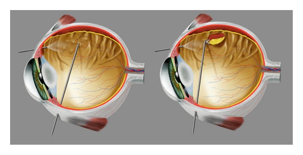
Introduction: Proliferative vitreoretinopathy (PVR) is a severe inflammatory complication of retinal detachment. Pathological epiretinal membranes grow on the retina surface leading to contraction, and surgery fails in 5% to 10% of the cases. We evaluated the expression of VEGF-A, Otx1, Otx2, Otx3, and p53 family members from PVR specimens to correlate their role in inducing or preventing the pathology.
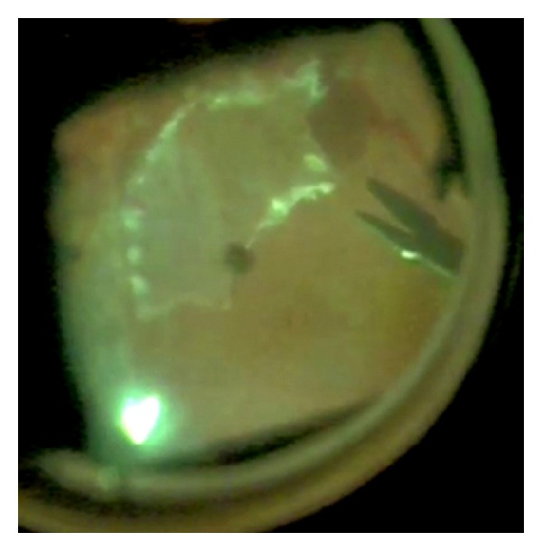
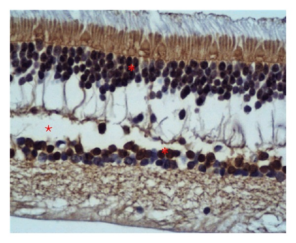
Methods: Twelve retinal samples were taken from patients affected by PVR during therapeutic retinectomies in vitreoretinal surgery. Gene expression was evaluated using quantitative real-time reverse transcriptase PCR analysis and immunohistochemistry, using four healthy human retinae as control.
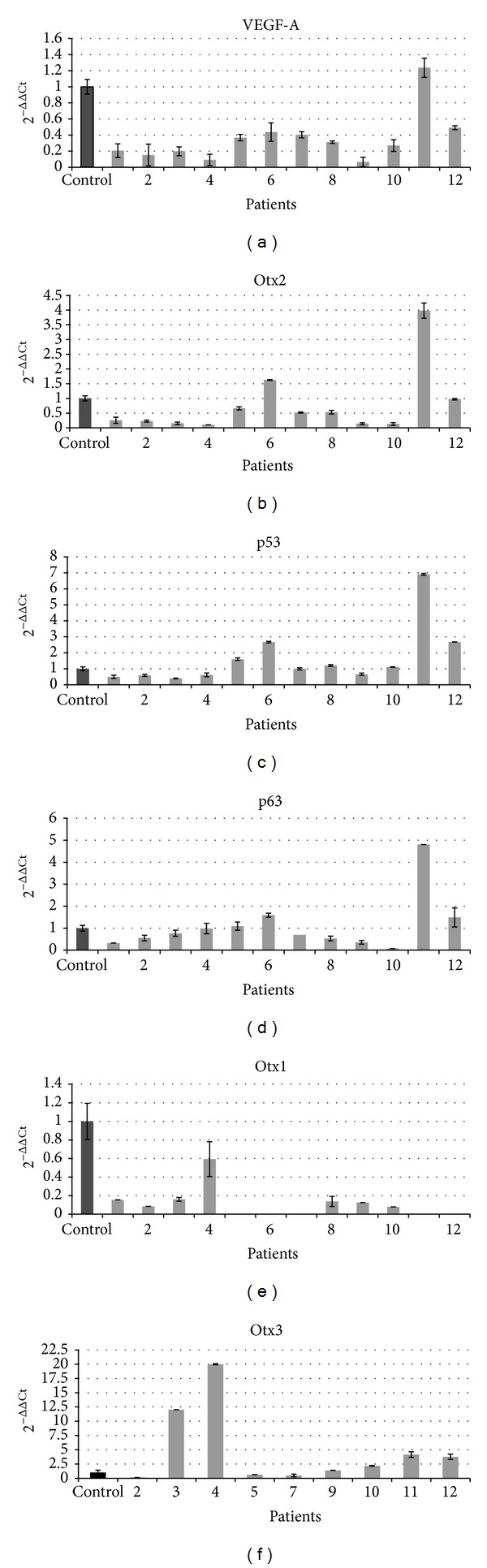
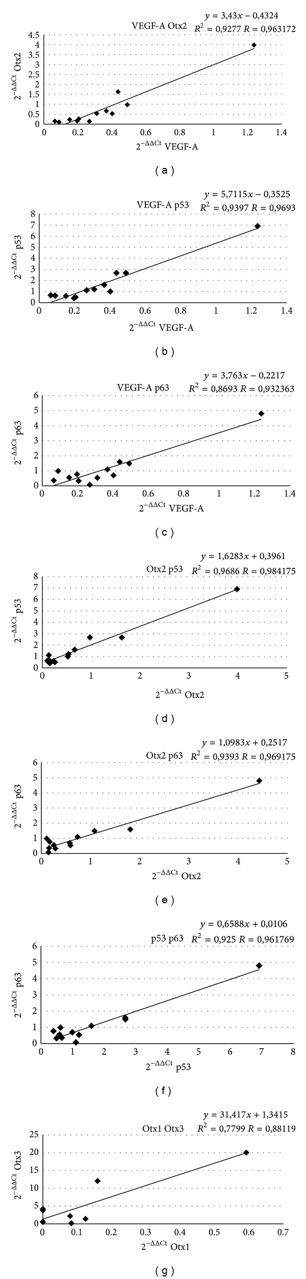
Result: Controls showed basal expression of all genes. PVR samples showed little or no expression of Otx1 and variable expression of VEGF-A, Otx2, Otx3, p53, and p63 genes. Significant correlation was found among VEGF-A, Otx2, p53, and p63 and between Otx1 and Otx3.
Conclusions: Otx homeobox, p53 family, and VEGF-A genes are expressed in PVR human retina. We individuated two possible pathways (VEGF-A, Otx2, p53, p63 and Otx1 and Otx3) involved in PVR progression that could influence in different manners the course of the pathology. Individuating the genetic pathways of PVR represents a novel approach to PVR therapies.
Figures
References
-
- Charles S, Calzada J. Vitreous Microsurgery. 5th edition. Philadelphia, Pa, USA: Lippincott Williams & Wilkins; 2010.
-
- Leiderman YI, Miller JW. Proliferative vitreoretinopathy: pathobiology and therapeutic targets. Seminars in Ophthalmology. 2009;24(2):62–69. - PubMed
-
- Ricker LJ, Kessels AGH, de Jager W, Hendrikse F, Kijlstra A, la Heij EC. Prediction of proliferative vitreoretinopathy after retinal detachment surgery: potential of biomarker profiling. The American Journal of Ophthalmology. 2012;154(2):347.e2–354.e2. - PubMed
-
- Symeonidis C, Papakonstantinou E, Androudi S, et al. Interleukin-6 and matrix metalloproteinase expression in the subretinal fluid during proliferative vitreoretinopathy: correlation with extent, duration of RRD and PVR grade. Cytokine. 2012;59(1):184–190. - PubMed
Publication types
MeSH terms
Substances
LinkOut - more resources
Full Text Sources
Other Literature Sources
Molecular Biology Databases
Research Materials
Miscellaneous
