

Successful treatment of melanocytoma associated choroidal neovascular membrane with intravitreal bevacizumab
- PMID: 24227972
- PMCID: PMC3809465
- DOI: 10.1016/j.sjopt.2012.12.002
Successful treatment of melanocytoma associated choroidal neovascular membrane with intravitreal bevacizumab
Abstract
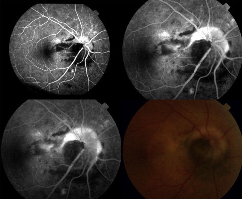
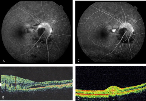
Melanocytoma of the optic disc is a benign melanocytic tumour that rarely causes visual impairment. We report a rare case of choroidal neovascularization (CNV) in association with optic disc melanocytoma and its response to intravitreal injection of the anti-vascular endothelial growth factor (VEGF), bevacizumab. The choroidal neovascular membrane regressed following a single intravitreal bevacizumab injection with formation of a scar. CNV associated with optic disc melanocytoma is rare. Intravitreal anti-VEGF treatment may be an effective treatment for CNV associated with optic disc melanocytoma.
Keywords: Anti-VEGF; Bevacizumab; Choroidal neovascular membrane; Melanocytoma; Optic disc.
Figures


References
-
- Bressler N.M., Bressler S.B., Fine S.L. Neovascular (exudative) age-related macular degeneration. In: Ryan S.J., editor. Retina. 4th ed. Elsevier Mosby; St. Louis: 2005. pp. 1075–1111.
-
- Chalam K.V., Shah V.A., Rappaport K.D. Choroidal neovascular membrane associated with melanocytoma of the optic nerve. Retina. 2006;26:703. - PubMed
-
- Joffe L., Shields J.A., Osher R.H., Gass J.D. Clinical and follow-up studies of melanocytomas of the optic disc. Ophthalmology. 1979;86:1067–1078. - PubMed
-
- Tran H.V., Bovey E.H., Uffer S., Zografos L. Peripapillary choroidal neovascularization associated with melanocytoma of the optic disc: a clinicopathologic case report. Graefes Arch Clin Exp Ophthalmol. 2006;244(10):1367–1369. - PubMed
LinkOut - more resources
Full Text Sources
Other Literature Sources
