. 2013 Jul;27(3):135-9.
doi: 10.1016/j.sjopt.2013.06.006.

Diffuse anterior retinoblastoma: A review

Affiliations

Diffuse anterior retinoblastoma: A review

Kristen P Jijelava et al. Saudi J Ophthalmol. 2013 Jul.

Abstract

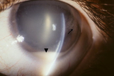
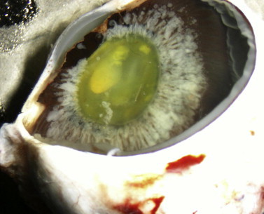
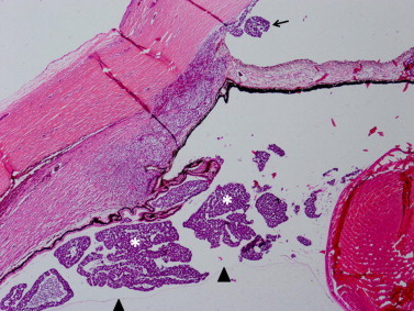
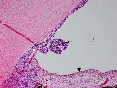
Diffuse anterior retinoblastoma is a rare variant of diffuse infiltrating retinoblastoma which occurs in up to 1-2% of cases of retinoblastoma. In diffuse anterior retinoblastoma there is a small focus of tumor in the peripheral retina from which free tumor cells enter the aqueous humor and implant on the ciliary body, iris, lens and trabecular meshwork. Patients most commonly present with pseudouveitis, pseudohypopyon and increased intraocular pressure. The differential diagnosis is broad and all of the reported cases relied upon aspirates from the aqueous humor in order to make the diagnosis prior to proceeding with treatment. Treatment involves enucleation and, depending upon the extent of the tumor, may require systemic chemotherapy or external beam radiation. This review summarizes the 7 previously reported cases of diffuse anterior retinoblastoma, discusses pathologic features, and addresses the challenges of early diagnosis and future directions.

Keywords: Anterior; Diffuse; Retinoblastoma; Uveitis.


Figures

Figure 1
Figure 1
Slit lamp appearance of diffuse anterior retinoblastoma. A white pseudohypopyon is present (arrowhead). There are also aggregates of white, fluffy tumor on the posterior corneal surface (arrow). The eye is otherwise white and quiet.
Figure 2
Figure 2
Gross appearance of enucleated eye with diffuse anterior retinoblastoma. There is white, fluffy material forming a corona around the lens. This represents tumor in the space between the anterior hyaloid face and ciliary body.
Figure 3
Figure 3
Diffuse anterior retinoblastoma in the posterior compartment. Tumor (asterisks) is present between the anterior hyaloid face (arrowheads) and the ciliary body. Tumor has percolated through the pupil and lodged in the peripheral anterior chamber (arrow). (Hematoxylin and eosin, 10X).
Figure 4
Figure 4
Diffuse anterior retinoblastoma in the anterior chamber. Tumor (arrow) is in the peripheral anterior chamber and plastered against the posterior corneal surface. There is neovascularization of the iris (arrowhead) present. Tumor has also infiltrated the trabecular meshwork (asterisk). (Hematoxylin and eosin, 100X).
None

Similar articles

Cited by

References

    1. Aziz H.A., LaSenna C.E., Vigoda M., Fernandes C., Feuer W., Aziz-Sultan M.A. Retinoblastoma treatment burden and economic cost: impact of age at diagnosis and selection of primary therapy. Clin Ophthalmol (Auckland, NZ) 2012;6:1601–1606. - PMC - PubMed
    1. Reis A.H., Vargas F.R., Lemos B. More epigenetic hits than meets the eye: microRNAs and genes associated with the tumorigenesis of retinoblastoma. Front Genet. 2012;3:284. - PMC - PubMed
    1. Shields C.L., Shields J.A. Diagnosis and management of retinoblastoma. Cancer Control. 2004;11(5):317–327. - PubMed
    1. Crosby M.B., Hubbard G.B., Gallie B.L., Grossniklaus H.E. Anterior diffuse retinoblastoma: mutational analysis and immunofluorescence staining. Arch Pathol Lab Med. 2009;133(8):215–218. - PMC - PubMed
    1. Shields C.L., Ghassemi F., Tuncer S., Thangappan A., Shields J.A. Clinical spectrum of diffuse infiltrating retinoblastoma in 34 consecutive eyes. Ophthalmology. 2008;115(12):2253–2258. - PubMed

LinkOut - more resources