. 2013 Jul;27(3):151-7.
doi: 10.1016/j.sjopt.2013.06.014.

Proton beam radiotherapy of uveal melanoma

Affiliations

Proton beam radiotherapy of uveal melanoma

Bertil Damato et al. Saudi J Ophthalmol. 2013 Jul.

Abstract

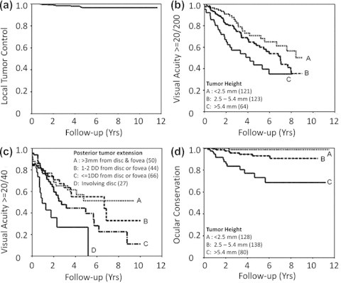
Proton beam radiotherapy of uveal melanoma can be administered as primary treatment, as salvage therapy for recurrent tumor, and as neoadjuvant therapy prior to surgical resection. The physical properties of proton beams make it possible to deliver high-doses of radiation to the tumor with relative sparing of adjacent tissues. This form of therapy is effective for a wider range of uveal melanoma than any other modality, providing exceptionally-high rates of local tumor control. This is particularly the case with diffuse iris melanomas, many of which are unresectable. The chances of survival, ocular conservation, visual preservation and avoidance of iatrogenic morbidity depend greatly on the tumor size, location and extent. When treating any side-effects and/or complications, it is helpful to consider whether these are the result of collateral damage or persistence of the irradiated tumor ('toxic tumor syndrome').

Keywords: Mortality; Neoadjuvant therapy; Proton beam radiotherapy; Recurrent tumor; Uveal melanoma.


Figures

Figure 1
Figure 1
Drawing showing tantalum markers sutured to the sclera. The distances from the tumor margins, the limbus and from each other are measured. (From B. Damato, Ocular Tumours: Diagnosis and Treatment, Butterworth Heineman, 2000).
Figure 2
Figure 2
Transilluminator for localizing posterior tantalum markers, designed by the first author (BD).
Figure 3
Figure 3
Proton beam treatment plan for a juxtapapillary choroidal melanoma in the left eye. The tumor had basal dimensions of 7.5 mm by 6.6 mm with a thickness of 0.9 mm. Three tantalum markers were inserted, using the optic disk as the ‘fourth marker’ (a). Proton beam diagram superimposed on a fundus photograph. Note the notch in the beam, designed to avoid collateral damage to the optic nerve (b). Three-dimensional diagram showing tumor, nerve, markers and beam (c). Vertical slice through eye showing minimal irradiation of the optic nerve (d).
Figure 4
Figure 4
Infero-nasal iris melanoma in the left eye of a 74-year-old woman. The tumor had basal dimensions of 4.4 mm by 2.2 mm with a thickness of 1.6 mm. Proton beam plan superimposed on the anterior-segment photograph (a). Horizontal section of the eye showing the penetration of the beam to the level of the ora (b).
Figure 5
Figure 5
Ocular outcomes of proton beam radiotherapy of choroidal melanoma. Local tumor control (a). Conservation of vision of 20/200 or better according to tumor height (b). Preservation of vision of 20/40 according to posterior tumor extension (c). Ocular conservation according to tumor height (d). (From Damato et al., Proton beam radiotherapy of choroidal melanoma: The Liverpool-Clatterbridge experience, International Journal of Radiation Oncology Biology Physics, Volume 62, Issue 5, 1 August 2005, Pages 1405–1411).
Figure 6
Figure 6
Visual acuity before proton beam radiotherapy of iris melanoma and at the latest known status. Each circle represents one case, and each line passing through this circle indicates an additional case. A = amblyopia; B = bullous keratopathy; C = cataract; G = glaucoma; H = hyphema; R = recurrence. (From Damato et al. Proton beam radiotherapy of iris melanoma, International Journal of Radiation Oncology Biology Physics, Volume 63, Issue 1, 1 September 2005, Pages 109–115).
None

References

    1. Groenewald C., Konstantinidis L., Damato B. Effects of radiotherapy on uveal melanomas and adjacent tissues. Eye (Lond) 2013;27(2):163–171. - PMC - PubMed
    1. Damato B., Kacperek A., Chopra M., Campbell I.R., Errington R.D. Proton beam radiotherapy of choroidal melanoma: the Liverpool-Clatterbridge experience. Int J Radiat Oncol Biol Phys. 2005;62(5):1405–1411. - PubMed
    1. Damato B., Kacperek A., Chopra M., Sheen M.A., Campbell I.R., Errington R.D. Proton beam radiotherapy of iris melanoma. Int J Radiat Oncol Biol Phys. 2005;63(1):109–115. - PubMed
    1. Konstantinidis L., Roberts D., Errington R.D., Kacperek A., Damato B. Whole anterior segment proton beam radiotherapy for diffuse iris melanoma. Br J Ophthalmol. 2013;97(4):471–474. - PubMed
    1. Gragoudas E.S. Proton beam irradiation of uveal melanomas: the first 30 years. The Weisenfeld Lecture. Invest Ophthalmol Vis Sci. 2006;47(11):4666–4673. - PubMed