

Orbital rhabdomyosarcomas: A review
- PMID: 24227982
- PMCID: PMC3770217
- DOI: 10.1016/j.sjopt.2013.06.004
Orbital rhabdomyosarcomas: A review
Abstract
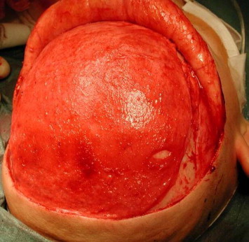
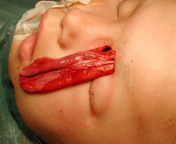
Rhabdomyosarcoma (RMS) is a highly malignant tumor and is one of the few life-threatening diseases that present first to the ophthalmologist. It is the most common soft-tissue sarcoma of the head and neck in childhood with 10% of all cases occurring in the orbit. RMS has been reported from birth to the seventh decade, with the majority of cases presenting in early childhood. Survival has changed drastically over the years, from 30% in the 1960's to 90% presently, with the advent of new diagnostic and therapeutic modalities. The purpose of this review is to provide a general overview of primary orbital RMS derived from a literature search of material published over the last 10 years, as well as to present two representative cases of patients that have been managed at our institute.
Keywords: AMORE; Brachytherapy; Eye; Genetics; Orbit; Pediatrics; Rhabdomyosarcoma; Tumor.
Figures

References
-
- Rootman J., editor. Neoplasia. Vol. 54. Lippincott Williams and Wilkins; Philadelphia: 2003. pp. 262–268. (Diseases of the orbit: a multidisciplinary approach).
-
- Shields J.A., Shields C.L. Rhabdomyosarcoma: review for the ophthalmologist. Surv Ophthalmol. 2003;48:39–57. - PubMed
-
- Turner J.H., Richmon J.D. Head and neck rhabdomyosarcoma: a critical analysis of population-based incidence and survival data. Otolaryngol Head Neck Surg. 2011;145:967–973. - PubMed
-
- Conneely M.F., Mafee M.F. Orbital rhabdomyosarcoma and simulating lesions. Neuroimaging Clin N Am. 2005;15:121–136. - PubMed
LinkOut - more resources
Full Text Sources
Other Literature Sources
