. 2013 Jul;27(3):203-8.
doi: 10.1016/j.sjopt.2013.07.004.

Secondary glaucoma as initial manifestation of uveal melanoma

Affiliations

Secondary glaucoma as initial manifestation of uveal melanoma

Ihab Saad Othman et al. Saudi J Ophthalmol. 2013 Jul.

Abstract

Purpose: Secondary glaucoma can be induced by a variety of local ocular problems. Intraocular tumors may initially present as secondary glaucoma.

Methods: 8 consecutive patients with secondary glaucoma were found to have uveal melanoma. Thorough examination included detailed history, fundus examination with scleral depression, B scan ultrasonography, and CT/MRI scanning techniques.

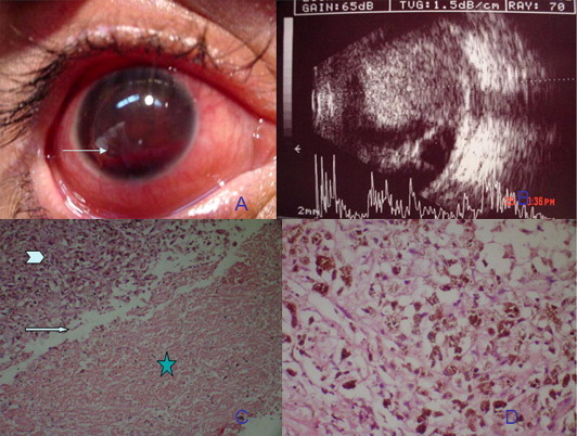
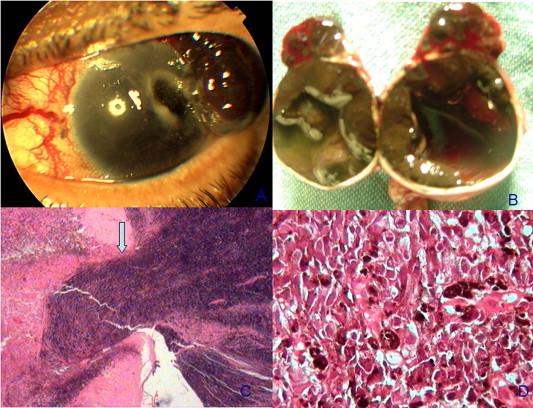
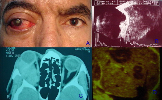
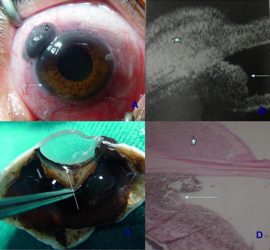
Results: A single case presented with spontaneous hyphema, two patients presented with secondary glaucoma, extraocular melanoma and metastases, a single case was found to have angle block by an iridociliary ring melanoma and 4 cases presented with neovascular glaucoma. Enucleation was necessary in all 8 cases.

Conclusions: General ophthalmologists should be aware of these rare initial manifestations of intraocular tumors as secondary glaucoma. Enucleation would be recommended in most cases of intraocular malignancy manifesting as secondary glaucoma. One should be extremely cautious in doing a penetrating surgery in such cases.

Keywords: Glaucoma; Melanoma; Neovascular glaucoma; Orbital melanoma.


Figures

Figure 1
Figure 1
(A) Anterior segment photography of case 1 showing hyphema filling the anterior chamber with level (arrow). (B) A and B scan ultrasonography showing a large choroidal melanoma occupying the posterior pole with associated retinal detachment. (C) Photomicrograph of the melanoma showing ghost cells in the hemorrhagic component (asterisk), sharp demarcation between melanoma cells (arrowhead) and ghost cells (arrow) (hematoxylin and eosin × 100). (D) Photomicrograph of epithelioid melanoma cells containing melanin pigment (Hematoxylin and eosin × 500).
Figure 2
Figure 2
(A) Anterior segment photography of a case of melanoma with extraocular extension inducing an epibulbar and corneal gutter. (B) Gross picture of the open globe following enucleation showing a diffuse intraocular melanoma occupying the choroid and ciliary body and extending on the surface of the globe. (C) Photomicrograph showing exit of melanoma cells from the trabeculectomy site. (Hematoxylin and eosin × 200). (D) Photomicrograph showing epithelioid melanoma cells. (Hematoxylin and eosin × 400).
Figure 3
Figure 3
(A) Facial photograph showing right axial proptosis with congested episcleral vessels OD. (B) A and B ultrasound scans showing a dome shaped choroidal melanoma with massive extrascleral extension. (C) Axial CT scan showing an isodense vitreous OD with a minimally elevated intraocular mass with massive retrobulbar extension OD. (D) B scan of the liver showing multiple echolucent masses within liver parenchyma denoting metastases.
Figure 4
Figure 4
(A) Photograph of the right eye of case – showing an epibulbar mass OD (asterisk), with a ring angle infiltration (arrow). (B) Ultrasound biomicroscopy of the same eye clearly delineating the epibulbar melanoma (asterisk) and the angle infiltration (arrow). (C) Gross photograph of the dissected specimen showing diffuse angle infiltration by melanoma cells. (arrow). (D) Photomicrograph of the enucleated globe showing correlation between UBM and histopathology showing the epibulbar melanoma 9asterisk and angle infiltration (arrow). (Hematoxylin and eosin × 100).
None

References

    1. Cunliffe I.A., Rennie I.G. Choroidal melanoma presenting as vitreous hemorrhage. Eye. 1993;7:711–713. - PubMed
    1. Gailloud C., Zografos L., Uffer S. Uveal melanomas and vitreous hemorrhage. Diagnosis and treatment. Klin Monatsbl Augenheilkd. 1991;198:165–170. - PubMed
    1. Haimovici R., Mukai S., Schachat A.P. Rhegmatogenous retinal detachment in eyes with uveal melanoma. Retina. 1996;16:488–496. - PubMed
    1. Damato B.E., Foulds W.S. Tumor-associated retinal pigment epitheliopathy. Eye. 1990:382–387. - PubMed
    1. Sneed S.R., Byrne S.F., Mieler W.F. Choroidal detachment associated with malignant choroidal tumors. Ophthalmology. 1991;98:963–970. - PubMed

LinkOut - more resources