

Diagnostic and therapeutic implications of a novel immunohistochemical panel detecting duodenal mucosal invasion by pancreatic ductal adenocarcinoma
- PMID: 24228110
- PMCID: PMC3816817
Diagnostic and therapeutic implications of a novel immunohistochemical panel detecting duodenal mucosal invasion by pancreatic ductal adenocarcinoma
Abstract
Background: We investigated a series of pancreaticoduodenectomy and duodenal biopsies with a panel of immunohistochemical markers to identify duodenal mucosal invasion by pancreatic ductal adenocarcinoma (PDAC), including markers of poor prognosis and targets of promising novel immunotherapies.
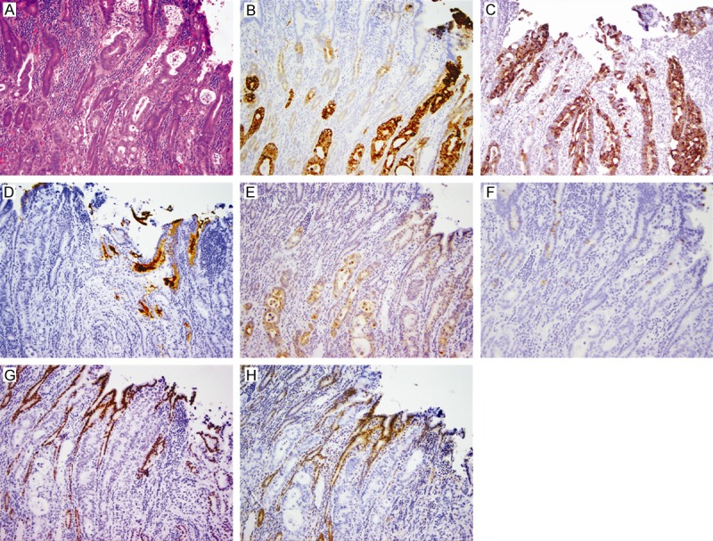
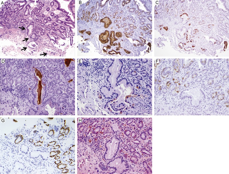
Materials and methods: Eighteen consecutive pancreaticoduodenectomy specimens with duodenal mucosal invasion by PDAC were examined for expression of MUC1, MUC4, MUC5AC, MUC6, mesothelin, MUC2, CDX2, and DPC4 on formalin-fixed, paraffin-embedded sections of duodenal-ampullary-pancreatic junctions. Expression of all but MUC6 was also assessed in duodenal biopsies from 12 patients with duodenal mucosal invasion by PDAC.
Results: The duodenal mucosa expressed MUC1 (crypts), MUC2 (goblet cells), MUC6 (Brunner glands), CDX2, and DPC4. PDACs in the duodenal mucosa from the resection (n=16-18) and biopsy (n=12) specimens were marked as follows: MUC1 100% (30/30), MUC4 83% (24/29), MUC5AC 83% (25/30), mesothelin 82% (23/28), MUC2 7% (2/30), and CDX2 36% (10/28). Loss of DPC4 expression was seen in 16 of 29 (55%) cases. Reactive mucosa adjacent to PDAC expressed MUC4, MUC5AC and mesothelin in 65% (17/26), 19% (5/27), and 19% (5/26) of cases, respectively. While MUC5AC and mesothelin had high diagnostic accuracy for detection of PDAC, MUC2, CDX2 and DPC4 expression demonstrated negative correlation with PDAC, with absent expression being highly specific for PDAC.
Conclusion: Immunohistochemical labeling for PDAC biomarkers may aid the diagnosis of PDAC in duodenal biopsy, especially in situations where diagnosis of a pancreatic mass is challenging.
Keywords: Pancreatic ductal adenocarcinoma; duodenal mucosal invasion; immunohistochemistry.
Figures

Similar articles
-
Diagnostic utility of mucin profile in fine-needle aspiration specimens of the pancreas: an immunohistochemical study with surgical pathology correlation.Cancer. 2006 Jun 25;108(3):186-97. doi: 10.1002/cncr.21913. Cancer. 2006. PMID: 16628655
-
Semiquantitative immunohistochemistry for mucin (MUC1, MUC2, MUC3, MUC4, MUC5AC, and MUC6) profiling of pancreatic ductal cell adenocarcinoma improves diagnostic and prognostic performance.Histopathology. 2016 Oct;69(4):582-91. doi: 10.1111/his.12994. Epub 2016 Jun 23. Histopathology. 2016. PMID: 27165582
-
Mucin expression in endoscopic ultrasound-guided fine-needle aspiration specimens is a useful prognostic factor in pancreatic ductal adenocarcinoma.Pancreas. 2015 Jul;44(5):728-34. doi: 10.1097/MPA.0000000000000362. Pancreas. 2015. PMID: 25906442 Free PMC article.
-
Well differentiation and intact Smad4 expression are specific features of groove pancreatic ductal adenocarcinomas.Pancreas. 2015 Apr;44(3):394-400. doi: 10.1097/MPA.0000000000000260. Pancreas. 2015. PMID: 25426619 Review.
-
Pathology of pancreatic ductal adenocarcinoma: facts, challenges and future developments.World J Gastroenterol. 2014 Oct 14;20(38):13833-41. doi: 10.3748/wjg.v20.i38.13833. World J Gastroenterol. 2014. PMID: 25320520 Free PMC article. Review.
Cited by
-
From Malignant Progression to Therapeutic Targeting: Current Insights of Mesothelin in Pancreatic Ductal Adenocarcinoma.Int J Mol Sci. 2020 Jun 6;21(11):4067. doi: 10.3390/ijms21114067. Int J Mol Sci. 2020. PMID: 32517181 Free PMC article. Review.
-
Diagnostic value of IMP3 in pancreatic cancer: a meta-analysis.Int J Clin Exp Med. 2015 Jul 15;8(7):10603-10. eCollection 2015. Int J Clin Exp Med. 2015. Retraction in: Int J Clin Exp Med. 2016 Jun 15;9(6):12417. PMID: 26379850 Free PMC article. Retracted.
-
Immunohistochemical Evaluation of the Expression of Specific Membrane Antigens in Patients with Pancreatic Ductal Adenocarcinoma.Cancers (Basel). 2023 Sep 15;15(18):4586. doi: 10.3390/cancers15184586. Cancers (Basel). 2023. PMID: 37760554 Free PMC article.
-
Quantitative assessment of the diagnostic role of mucin family members in pancreatic cancer: a meta-analysis.Ann Transl Med. 2021 Feb;9(3):192. doi: 10.21037/atm-20-5606. Ann Transl Med. 2021. PMID: 33708819 Free PMC article.
-
Diagnostic value of mesothelinin pancreatic cancer: a meta-analysis.Int J Clin Exp Med. 2014 Nov 15;7(11):4000-7. eCollection 2014. Int J Clin Exp Med. 2014. PMID: 25550908 Free PMC article.
References
-
- Surveillance Epidemiology and End Results. SEER Fact Sheets. 2012.
-
- Sohn TA, Yeo CJ, Cameron JL, Koniaris L, Kaushal S, Abrams RA, Sauter PK, Coleman J, Hruban RH, Lillemoe KD. Resected adenocarcinoma of the pancreas-616 patients: results, outcomes, and prognostic indicators. J Gastrointest Surg. 2000;4:567–579. - PubMed
-
- Yeo CJ, Abrams RA, Grochow LB, Sohn TA, Ord SE, Hruban RH, Zahurak ML, Dooley WC, Coleman J, Sauter PK, Pitt HA, Lillemoe KD, Cameron JL. Pancreaticoduodenectomy for pancreatic adenocarcinoma: postoperative adjuvant chemoradiation improves survival. A prospective, single-institution experience. Ann Surg. 1997;225:621–633. discussion 633-626. - PMC - PubMed
Publication types
MeSH terms
Substances
Grants and funding
LinkOut - more resources
Full Text Sources
Medical
Research Materials
Miscellaneous