Nasal lobular capillary hemangioma
- PMID: 24228209
- PMCID: PMC3814904
- DOI: 10.4103/2156-7514.119134
Nasal lobular capillary hemangioma
Abstract
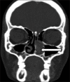
Nasal lobular capillary hemangioma is a rare benign tumor of the paranasal sinuses. This lesion is believed to grow rapidly in size over time. The exact etiopathogenesis is still a dilemma. We discuss a case of nasal lobular capillary hemangioma presenting with a history of epistaxis. Contrast enhanced computed tomography of paranasal sinuses revealed an intensely enhancing soft-tissue mass in the left nasal cavity and left middle and inferior meati with no obvious bony remodeling or destruction. We present imaging and pathologic features of nasal lobular capillary hemangioma and differentiate it from other entities like nasal angiofibroma.
Keywords: Kiesselbach plexus; Nasal lobular capillary hemangioma; paranasal sinuses; pyogenic granuloma.
Conflict of interest statement
Figures


References
-
- Ozcan C, Apa DD, Görür K. Pediatric lobular capillary hemangioma of the nasal cavity. Eur Arch Otorhinolaryngol. 2004;261:449–51. - PubMed
-
- Pagliai KA, Cohen BA. Pyogenic granuloma in children. Pediatr Dermatol. 2004;21:10–3. - PubMed
-
- Poncet A, Dor L, Botyromycose Humaine Rev Chir (Paris) 1897;18:996.
-
- Mills SE, Cooper PH, Fechner RE. Lobular capillary hemangioma: The underlying lesion of pyogenic granuloma. A study of 73 cases from the oral and nasal mucous membranes. Am J Surg Pathol. 1980;4:470–9. - PubMed
-
- Miller FR, D’Agostino MA, Schlack K. Lobular capillary hemangioma of the nasal cavity. Otolaryngol Head Neck Surg. 1999;120:783–4. - PubMed
LinkOut - more resources
Full Text Sources
Other Literature Sources

