. 2013 Sep 30:3:41.
doi: 10.4103/2156-7514.119139. eCollection 2013.

Multispectral Photoacoustic Imaging of Prostate Cancer: Preliminary Ex-vivo Results

Affiliations

Multispectral Photoacoustic Imaging of Prostate Cancer: Preliminary Ex-vivo Results

Vikram S Dogra et al. J Clin Imaging Sci. .

Abstract

Objective: The objective of this study is to validate if ex-vivo multispectral photoacoustic (PA) imaging can differentiate between malignant prostate tissue, benign prostatic hyperplasia (BPH), and normal human prostate tissue.

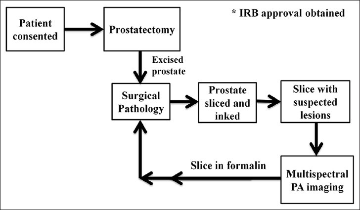
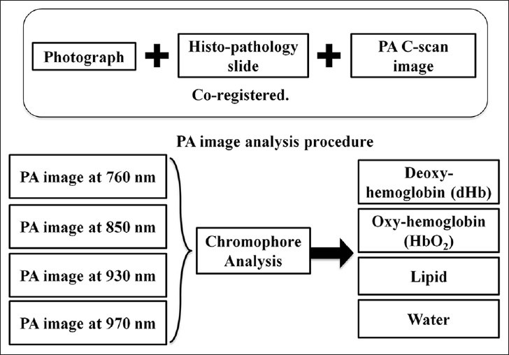
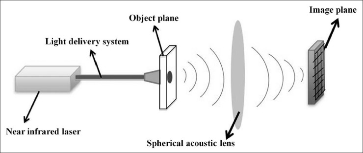
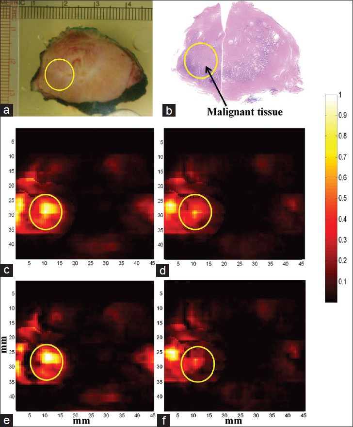
Materials and methods: Institutional Review Board's approval was obtained for this study. A total of 30 patients undergoing prostatectomy for biopsy-confirmed prostate cancer were included in this study with informed consent. Multispectral PA imaging was performed on surgically excised prostate tissue and chromophore images that represent optical absorption of deoxyhemoglobin (dHb), oxyhemoglobin (HbO2), lipid, and water were reconstructed. After the imaging procedure is completed, malignant prostate, BPH and normal prostate regions were marked by the genitourinary pathologist on histopathology slides and digital images of marked histopathology slides were obtained. The histopathology images were co-registered with chromophore images. Region of interest (ROI) corresponding to malignant prostate, BPH and normal prostate were defined on the chromophore images. Pixel values within each ROI were then averaged to determine mean intensities of dHb, HbO2, lipid, and water.

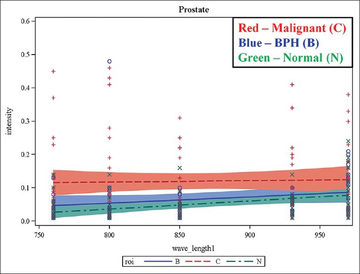
Results: Our preliminary results show that there is statistically significant difference in mean intensity of dHb (P < 0.0001) and lipid (P = 0.0251) between malignant prostate and normal prostate tissue. There was difference in mean intensity of dHb (P < 0.0001) between malignant prostate and BPH. Sensitivity, specificity, positive predictive value, and negative predictive value of our imaging system were found to be 81.3%, 96.2%, 92.9% and 89.3% respectively.

Conclusion: Our preliminary results of ex-vivo human prostate study suggest that multispectral PA imaging can differentiate between malignant prostate, BPH and normal prostate tissue.

Keywords: Multispectral; photoacoustic; prostate.


Conflict of interest statement

Conflict of Interest: None declared.

Figures

Figure 1
Figure 1
Tissue acquisition and handling protocol for photoacoustic imaging of ex-vivo prostate specimens.
Figure 2
Figure 2
Photoacoustic (PA) image analysis procedure. Chromophore analysis was performed on the acquired multispectral composite PA images to extract individual optical absorption maps of dHb, HbO2, lipid and water. Each PA image is co-registered with the photograph of gross prostate tissue and histopathology for further evaluation.
Figure 3
Figure 3
Results of generalized estimated equation model demonstrating the potential of multispectral photoacoustic imaging in differentiating normal, BPH and malignant prostate tissue. The plots show mean intensity values against incident laser wavelength. The mean intensity of malignant prostate was found to be higher compared with that normal and BPH at all the wavelengths.
Figure 4
Figure 4
Illustration of photoacoustic (PA) effect. When a tissue is exposed to low energy pulsed laser beam for a very short duration, optical absorption in the tissue takes place followed by localized heating and rapid thermal expansion generating acoustic waves. Since the acoustic waves are generated due to laser exposure, they are commonly called PA waves.
Figure 5
Figure 5
Differences between C-scan and B-scan image formation. B-scan image depicts sagittal or transverse planes in the body where as a C-scan image depicts information from coronal plane in the body. Reproduced with permission from “Basics and Clinical Applications of Photoacoustic Imaging,” Ultrasound Clinics, Vol. 4, Issue 3: 403-429, July 2009.
Figure 6
Figure 6
Acoustic lens focusing. Photoacoustic signals generated from the tissue (object plane) are focused onto an ultrasound sensor array placed in the image plane, enabling real-time high speed data acquisition. Reproduced with permission from “Photoacoustic Imaging: Opening New Frontiers in Medical Imaging”. J Clin Imaging Sci 2011, 1:24.
Figure 7
Figure 7
Multispectral photoacoustic (PA) imaging of prostate. PA images are acquired at multiple laser wavelengths. Each wavelength image is a composite image of all the tissue constituents such as deoxy-hemoglobin (dHb), oxy-hemoglobin (HbO2), lipid and water. Chromophore analysis was performed to extract PA images showing absorption of individual constituents from the multi-wavelength images. All the PA images are co-registered with histopathology and photograph of the gross specimen. (a) Photograph of gross prostate specimen (b) Histopathology of prostate with malignant region encircled. (c) Composite PA image acquired at 760 nm wavelength (d) Composite PA image acquired at 850 nm wavelength (e) PA image showing absorption of dHb (f) PA image showing absorption of HbO2. Higher absorption of dHb was seen in the region of interest corresponding to malignant prostate tissue compared to HbO2.

Similar articles

Cited by

References

    1. Eheman C, Henley SJ, Ballard-Barbash R, Jacobs EJ, Schymura MJ, Noone AM, et al. Annual Report to the nation on the status of cancer, 1975-2008, featuring cancers associated with excess weight and lack of sufficient physical activity. Cancer. 2012;118:2338–66. - PMC - PubMed
    1. Cancer Statistics. National Cancer Institute. 2012. [Last accessed on 2013 Apr 24]. Available from: http://www.cancer.gov/statistics .
    1. Carter HB, Hamper UM, Sheth S, Sanders RC, Epstein JI, Walsh PC. Evaluation of transrectal ultrasound in the early detection of prostate cancer. J Urol. 1989;142:1008–10. - PubMed
    1. Rao NA, Lai D, Bhatt S, Arnold SC, Chinni B, Dogra VS. Acoustic lens characterization for ultrasound and photoacoustic C-scan imaging modalities. Conf Proc IEEE Eng Med Biol Soc 2008. 2008:2177–80. - PubMed
    1. Valluru KS, Chinni BK, Rao NA, Bhatt S, Akata D, Dogra VS. Development of a c-scan photoacoustic imaging probe for prostate cancer detection. Proc SPIE. 2011;7968 79680C1-7.

LinkOut - more resources