. 2013 Sep 24;2(1):12.
doi: 10.1186/2046-2530-2-12.

Ciliogenesis and cerebrospinal fluid flow in the developing Xenopus brain are regulated by foxj1

Affiliations

Ciliogenesis and cerebrospinal fluid flow in the developing Xenopus brain are regulated by foxj1

Cathrin Hagenlocher et al. Cilia. .

Abstract

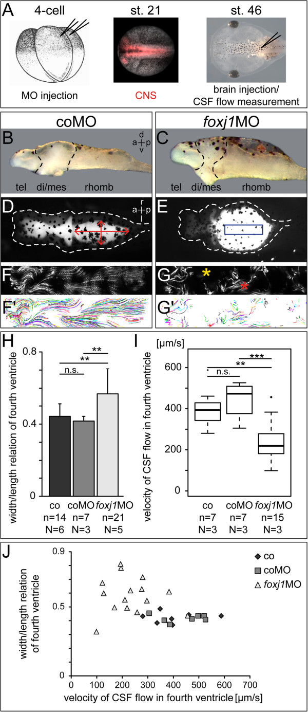
Background: Circulation of cerebrospinal fluid (CSF) through the ventricular system is driven by motile cilia on ependymal cells of the brain. Disturbed ciliary motility induces the formation of hydrocephalus, a pathological accumulation of CSF resulting in ventricle dilatation and increased intracranial pressure. The mechanism by which loss of motile cilia causes hydrocephalus has not been elucidated. The aim of this study was: (1) to provide a detailed account of the development of ciliation in the brain of the African clawed frog Xenopus laevis; and (2) to analyze the relevance of ependymal cilia motility for CSF circulation and brain ventricle morphogenesis in Xenopus.

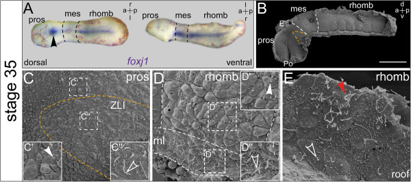
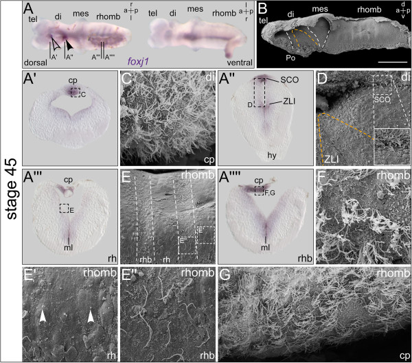
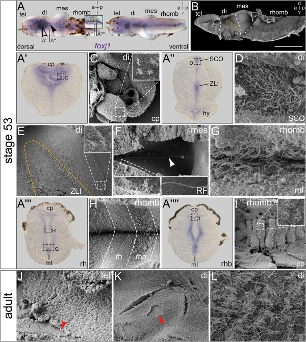
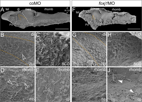
Methods: Gene expression analysis of foxj1, the bona fide marker for motile cilia, was used to identify potentially ciliated regions in the developing central nervous system (CNS) of the tadpole. Scanning electron microscopy (SEM) was used to reveal the distribution of mono- and multiciliated cells during successive stages of brain morphogenesis, which was functionally assessed by bead injection and video microscopy of ventricular CSF flow. An antisense morpholino oligonucleotide (MO)-mediated gene knock-down that targeted foxj1 in the CNS was applied to assess the role of motile cilia in the ventricles.

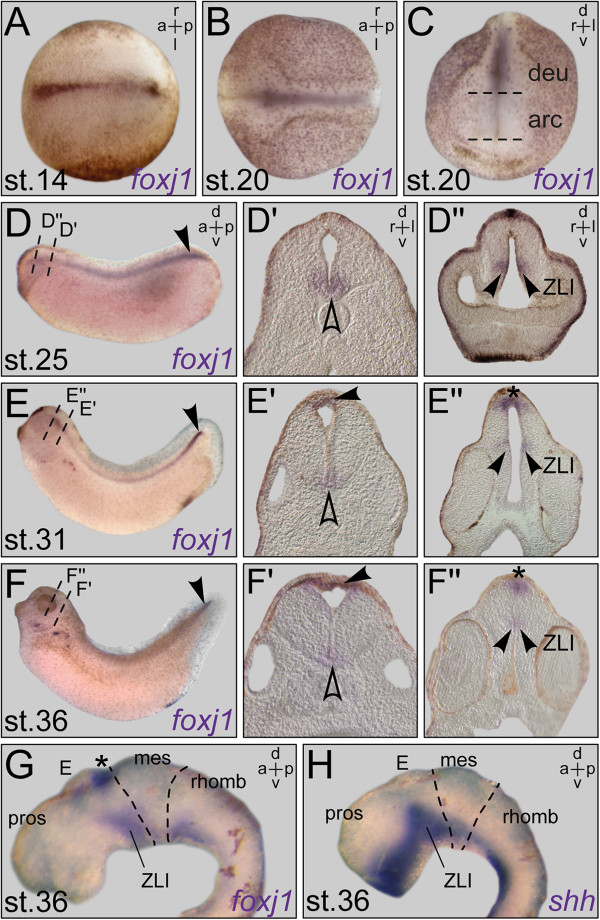
Results: RNA transcripts of foxj1 in the CNS were found from neurula stages onwards. Following neural tube closure, foxj1 expression was seen in distinct ventricular regions such as the zona limitans intrathalamica (ZLI), subcommissural organ (SCO), floor plate, choroid plexus (CP), and rhombomere boundaries. In all areas, expression of foxj1 preceded the outgrowth of monocilia and the subsequent switch to multiciliated ependymal cells. Cilia were absent in foxj1 morphants, causing impaired CSF flow and fourth ventricle hydrocephalus in tadpole-stage embryos.

Conclusions: Motile ependymal cilia are important organelles in the Xenopus CNS, as they are essential for the circulation of CSF and maintenance of homeostatic fluid pressure. The Xenopus CNS ventricles might serve as a novel model system for the analysis of human ciliary genes whose deficiency cause hydrocephalus.


Figures

Figure 1
Figure 1
Xenopus tadpoles express foxj1 in distinct regions of the developing CNS. Whole-mount in situ hybridization of staged embryos and explanted brains. (A) Expression at early neurula stage (dorsal (d) view). (B, C) Expression at late neurula stage in epidermis and floor plate, in dorsal (B) and frontal view (C). (D) Staining in the floor plate at stage 25. Note the higher expression level at the ampulla terminalis (arrowhead). (D?, D?) Transversal sections, levels indicated in (D), revealed staining in the ventral (v) midline (outlined arrowhead) (D?) and the zona limitans intrathalamica (ZLI; arrowheads) (D?). (E, F) Expression at stage 31 (E) and stage 36 (F) in the nephrostomes, the spinal cord, and the ampulla terminalis (arrowheads). (E?, E?, F?, F?) Histological sections as indicated in (E, F) highlighted expression in the rhombencephalic roof (arrowheads in E?, F?), the ventral midline (outlined arrowheads in E?, F?) as well as in the subcommissural organ (SCO; asterisk in E?, F?, G) and the ZLI (arrowheads in E?, F?). (G, H) Brain explants at stage 36 after in situ hybridization with antisense probes for foxj1(G) and shh(H) in side view showed co-localization of mRNA expression in the ventral midline and the ZLI. a = anterior; arc = archencephalon; deu = deuterencephalon; l = left; mes = mesencephalon; p = posterior; pros = prosencephalon; rhomb = rhombencephalon; r = right; st. = stage.
Figure 2
Figure 2
foxj1 expression correlates with elongation of monocilia and the emergence of multiple cilia. In situ hybridization and scanning electron microscopy (SEM) on explanted brains at stage 35. (A) Explant shown in dorsal (d) and ventral (v) view. Strong expression in the ventral midline and the subcommissural organ (SCO; arrowhead). (B) SEM picture of brain explant dissected sagittally with view onto the ventricular surface, the zona limitans intrathalamica (ZLI) is delimited with orange dashed line and boundaries between brain regions are indicated by white dashed lines. Bar represents 200 ?m. (C) Overview and enlargements show short primary cilia on cells in the prosencephalic region (arrowhead in (C?)), and elongated monocilia on cells within the ZLI (outlined arrowhead in (C?)). (D) Close-up view onto the ventral aspect of a single hindbrain rhombomere with indicated boundaries and midline (ml). Enlargements showing short cilia on the rhombomere (arrowhead in (D?)) as well as elongated cilia in the ventral ml (outlined arrowhead in (D?)). (E) Close-up view onto the rhombencephalon (rhomb) roof with elongated monocilia (outlined arrowhead) and first MCCs (arrowhead). a = anterior; E = epiphysis; l = left; mes = mesencephalon; p = posterior; Po = preoptic region; pros = prosencephalon; r = right.
Figure 3
Figure 3
Additional foxj1 expression domains identify regions with emerging ciliation. In situ hybridization and scanning electron microscopy (SEM) on explanted brains at stage 45. (A) Explant shown in dorsal (d) and ventral (v) view. Expression in the ventral midline, rhombomere boundaries, subcommissural organ (SCO, arrowhead), and the choroid plexus (cp; outlined arrowhead). (B) SEM picture of brain explant dissected sagittally with view onto the ventricular surface, the zona limitans intrathalamica (ZLI) is delimited with orange dashed line and boundaries between brain regions are indicated by white dashed lines. Bar represents 200 ?m. (A?-A??) Transversal histological sections as indicated in (A), dorsal side up, and SEM pictures (C-G) of corresponding regions as indicated in the sections, respectively. (C) MCCs on cp invaginating from the diencephalon roof. (D) Close-up view onto the ZLI region; enlargement showing ciliated structure of the SCO. (E) Close-up view revealing metamerical organization of the rhombencephalon (rhomb). Rhombomeres (rh) and rh boundaries (rhb) indicated by dashed lines. (E?) Enlargement of the rh region; arrowheads pointing to short primary cilia. (E?) Enlargement of the rhb with cells bearing elongated monocilia. (F, G) Close-up views onto the fourth ventricle cp showing MCCs. a = anterior; di = diencephalon; hy = hypophysis; l = left; mes = mesencephalon; ml = midline; p = posterior; Po = preoptic region; r = right; tel = telencephalon.
Figure 4
Figure 4
Continued foxj1 expression and ciliogenesis in the premetamorphic and adult brain of Xenopus. In situ hybridization and scanning electron microscopy (SEM) on explanted brains at stage 53 and SEM on adult brains. (A) Explant shown in dorsal (d) and ventral (v) view. Expression in the ventral midline, subcommissural organ (SCO, arrowhead), choroid plexus (cp; outlined arrowhead), and ventricular regions. Inset in (A) focuses on expression in rhombomere boundaries (rhb) after removal of the pigmented rhombencephalon (rhomb) roof. (B) SEM picture of brain explant dissected sagittally with view onto the ventricular surface, the zona limitans intrathalamica (ZLI) is delimited with orange dashed line and boundaries between brain regions are indicated by white dashed lines. Bar represents 500 ?m. (A?-A??) Transversal histological sections as indicated in (A), dorsal side up. (C-I) SEM pictures corresponding to regions indicated in (A?-A??), respectively. (C) Close-up view of third ventricle cp; inset showing enlarged view of MCCs. (D) Top view onto ventricular surface of SCO of a frontally bisected specimen. (E) Overview of the diencephalon (di) region; inset showing MCCs emerging at the di-/mesencephalic boundary, similar to those of the ZLI. (F-I) Frontally bisected specimens. (F) View into the dorsal ventricular lumen of the mesencephalon (mes) with Reissner?s fiber (RF, arrowhead) spanning the ventricle. Left inset: enlargement of mes MCCs. Right inset: enlargement of RF. (G) View onto the ventral midline (ml). (H) Ventral part of the rhomb with rhombomeres (rh) and rhb as indicated. (I) cp of the rhomb roof with MCCs. (J-L) SEM pictures of sagittally bisected adult brain in the telencephalon (tel; (J)) and di region (K, L) showing a multiciliated ependymal monolayer. Arrows indicate sites of ependyma detachment due to mechanical stress during processing. a = anterior; hy = hypophysis; l = left; p = posterior; r = right.
Figure 5
Figure 5
Loss of function of foxj1 in the central nervous system induces hydrocephalus. (A) Timeline of experimental process. (B, D, F, F?) Control morpholino (coMO) -injected specimen. (C, E, G, G?) foxj1 morpholino (foxj1MO) -injected specimen. (B, C) Brain explants of stage 46 tadpoles, side view. (D, E) Dorsal (d) view of tadpole heads at stage 46; ventricular system visualized by injection of a fluorescent dye. Note the obstruction of the ventricular system and the hydrocephalic hindbrain ventricle in foxj1MO -injected embryos in (E). (F, G) Z-projection of video-tracked fluorescent beads. (F?, G?) Display of statistically analyzed bead trajectories. (H) Statistical analysis of fourth ventricle width to length relation, measured as indicated in (D). 0 = linear ventricle, 1 = perfect circle; error bars represent SD. (I) Statistical analysis of CSF flow velocity in the fourth ventricle. Region of interest chosen for flow measurement as indicated by blue box in (E). (J) Correlation of statistical analyses in (H) and (I). a = anterior; CNS = central nervous system; co = control; CSF = cerebrospinal fluid; di = diencephalon; l = left; mes = mesencephalon; n = number of specimens; N = number of experiments; p = posterior; r = right, rhomb = rhombencephalon; tel = telencephalon; v = ventral.
Figure 6
Figure 6
Reduced CSF flow velocity correlates with defects in ciliogenesis. Scanning electron microscopy (SEM) of dissected brain explants at stage 46. (A-E) Control morpholino (coMO) -injected specimen. (F-J)foxj1 morpholino (foxj1MO) -injected specimen. (A,F) SEM pictures of brain explants dissected sagittally with view on the ventricular surface, the zona limitans intrathalamica (ZLI) is delimited with orange dashed line and boundaries between brain regions indicated by white dashed lines. Bars represent 200 ?m. (B,G) View onto the ZLI in the diencephalon (di). Note the loss of cilia on the ZLI in foxj1 morphants (G). (C,H) Close-up views onto the subcommissural organ (SCO) in frontally dissected specimens. (D,I) Close-up views onto the rhombencephalon in sagittally dissected specimens, depicting the rhombomere boundaries. Outlined arrows in (D) point to elongated monocilia. Note the absence of cilia in (I). (E,J) Close-up view onto the choroid plexus (cp) of the hindbrain roof. Arrowheads in (J) point to the few remaining MCCs in foxj1 morphants. a = anterior; d = dorsal; mes = mesencephalon; tel = telencephalon; rhomb = rhombencephalon; Po = preoptic region; p = posterior; rhb = rhombomere boundary; v = ventral.

References

    1. Brown PD, Davies SL, Speake T, Millar ID. Molecular mechanisms of cerebrospinal fluid production. Neuroscience. 2004;129:957–970. - PMC - PubMed
    1. Kapoor KG, Katz SE, Grzybowski DM, Lubow M. Cerebrospinal fluid outflow: an evolving perspective. Brain Res Bull. 2008;77:327–334. doi: 10.1016/j.brainresbull.2008.08.009. - DOI - PubMed
    1. Del Bigio MR. Ependymal cells: biology and pathology. Acta Neuropathol. 2010;119:55–73. doi: 10.1007/s00401-009-0624-y. - DOI - PubMed
    1. Sawamoto K, Wichterle H, Gonzalez-Perez O, Cholfin J, Yamada M, Spassky N, Murcia N, Garcia-Verdugo J, Marin O, Rubenstein J, Tessier-Lavigne M, Okano H, Alvarez-Buylla A. New neurons follow the flow of cerebrospinal fluid in the adult brain. Science. 2006;311:629–632. doi: 10.1126/science.1119133. - DOI - PubMed
    1. Iba?ez-Tallon I, Pagenstecher A, Fliegauf M, Olbrich H, Kispert A, Ketelsen UP, North A, Heintz N, Omran H. Dysfunction of axonemal dynein heavy chain Mdnah5 inhibits ependymal flow and reveals a novel mechanism for hydrocephalus formation. Hum Mol Genet. 2004;13:2133–2141. doi: 10.1093/hmg/ddh219. - DOI - PubMed