Clinical Trial
. 2013 Dec;123(12):5310-8.
doi: 10.1172/JCI70314. Epub 2013 Nov 15.

Human Treg responses allow sustained recombinant adeno-associated virus-mediated transgene expression

Clinical Trial

Human Treg responses allow sustained recombinant adeno-associated virus-mediated transgene expression

Christian Mueller et al. J Clin Invest. 2013 Dec.

Abstract

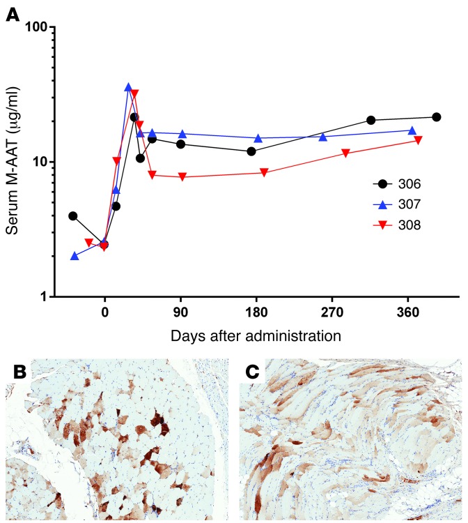
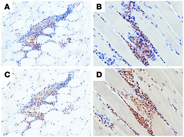
Recombinant adeno-associated virus (rAAV) vectors have shown promise for the treatment of several diseases; however, immune-mediated elimination of transduced cells has been suggested to limit and account for a loss of efficacy. To determine whether rAAV vector expression can persist long term, we administered rAAV vectors expressing normal, M-type α-1 antitrypsin (M-AAT) to AAT-deficient subjects at various doses by multiple i.m. injections. M-specific AAT expression was observed in all subjects in a dose-dependent manner and was sustained for more than 1 year in the absence of immune suppression. Muscle biopsies at 1 year had sustained AAT expression and a reduction of inflammatory cells compared with 3 month biopsies. Deep sequencing of the TCR Vβ region from muscle biopsies demonstrated a limited number of T cell clones that emerged at 3 months after vector administration and persisted for 1 year. In situ immunophenotyping revealed a substantial Treg population in muscle biopsy samples containing AAT-expressing myofibers. Approximately 10% of all T cells in muscle were natural Tregs, which were activated in response to AAV capsid. These results suggest that i.m. delivery of rAAV type 1-AAT (rAAV1-AAT) induces a T regulatory response that allows ongoing transgene expression and indicates that immunomodulatory treatments may not be necessary for rAAV-mediated gene therapy.


Figures

Figure 1
Figure 1. M-AAT expression in skeletal muscle from AAT-deficient human subjects ≥12 months after i.m. injection of rAAV1-hAAT.
All subjects in this dose cohort received 6.0 × 10¹² vg/kg. The body weight maximum was 90 kg, thus individual doses ranged up to 5.4 × 10¹⁴ total vg. The therapeutic target was 572 μg/ml. (A) Serum AAT levels detected using a PiM-specific ELISA in subjects 306, 307, and 308. (B and C) Muscle immunohistochemistry staining for hAAT. Specimens were biopsied from each individual 1 year after rAAV administration and stained for the presence of AAT. Sections show granular reactivity in individual myofibers on cross-section. Immunohistochemistry staining for hAAT in a normal, noninjected muscle is shown in Supplemental Figure 4. Original magnification, ×5.
Figure 2
Figure 2. Persistence of lymphocytic infiltrates in muscle more than 1 year after administration (A) Immunohistochemistry showing a nidus of infiltration with CD3+ T cells.
(B) CD4-immunoreactive T cells comprise a substantial subset of the total lymphocytic infiltrate. (C) CD8-immunoreactive T cells are also present in the infiltrate. (D) CD68+ macrophages are present within and around the lymphocytic infiltrates. Original magnification, ×10. (E) Representative time course of IFN-γ ELISPOT (subject 307) responses to pools of AAV1 capsid peptides or controls. PBMCs were obtained at screening, baseline, and 1, 2, 3, and approximately 18 months after vector administration and were stimulated with one of three pools (AC) of AAV1 capsid peptides (15-mers overlapped by 10 amino acids) or with a positive control peptide pool (CEF). SFC, spot-forming cells. Positive responses to AAV1 capsid peptides are indicated by *.
Figure 3
Figure 3. TCR β-chain deep sequencing analysis in subject 306.
(A) Frequency of T cell clones with a frequency greater than 0.005% that were present in blood only after gene transfer and in injected muscle tissue after 3 months and 1 year of vector administration. (B) Frequency of T cell clones arranged by specific Vβ chain in muscle biopsies (B) at 3 months and (C) after 1 year of vector administration. Colors in each stacked bar indicate unique T cell clones.
Figure 4
Figure 4. In situ detection of Tregs at 12 months after vector administration.
(AC) Formalin-fixed, paraffin-embedded muscle biopsies from patients were stained with antibodies specific for the cell surface markers CD4 and CD25 and the transcription factor FOXP3. Original magnification, ×10. (D) The merged image shows the presence of Tregs in muscle tissue. Flash frozen muscle sections were also stained with DAPI (blue), CD4 (red), and FOXP3 (yellow) for higher resolution of FOXP3 localization. E and F show higher-magnification images of FOXP3+CD4+ cells in the muscle biopsies. Original magnification, ×10 (AD); ×40 (E and F). (GI) Analysis of the FOXP3 TSDR. Epigenetic detection was used to quantify total (G) CD3+ cells and (H) Tregs in the muscle biopsies of patients 304 and 308 and (I) the proportion of muscle T cell infiltrates that that were Tregs.
Figure 5
Figure 5. Antigen-specific activation of Tregs.
PBMCs were stimulated with AAV, AAT peptide pools, or 1 μg/ml CEFT peptide. Cells were harvested at 48 hours after activation and gated for live CD4+, FOXP3+, and Helios+ cells and then subgated for activation markers OX40+ and CD25+. Lymphocytes were gated on forward and side scatter gates. Live CD4+ T cells were subgated for analysis of specific subsets as follows. Tregs were gated by coexpression of the transcription factors FOXP3 and Helios. Conventional T cells were gated as CD4+FOXP3Helios. All subsets were then analyzed for expression of CD25 and OX40 as indicators of antigen-specific activation. The data are plotted as activation above CD4+ CEFT stimulation. Data are shown as an average for all 8 samples ± SEM.
Figure 6
Figure 6. In situ detection of intact AAV1 capsid in the muscle at 3 and 12 months after vector administration.
Muscle biopsies at (A) 3 months and (B) 12 months after administration of the vector were stained with DAPI (blue, top left) and an antibody specific for AAV1 intact capsid (red, bottom left). The top right image of each panel shows a DIC image, and the bottom right image of each panel is a merged image. Original magnification, ×40.
Figure 7
Figure 7. Representative in situ detection of PD-1 and PD-L1 in muscle lymphocytic infiltrates.
Muscle biopsies were stained for (A and B) PD-1 or (C and D) PD-L1. Sections show positive staining for PD-1 and PD-L1 in lymphocyte infiltrates. Original magnification, ×10 (A and C); ×20 (B and D).

References

    1. Maguire AM, et al. Safety and efficacy of gene transfer for Leber’s congenital amaurosis. N Engl J Med. 2008;358(21):2240–2248. doi: 10.1056/NEJMoa0802315. - DOI - PMC - PubMed
    1. Hauswirth WW, et al. Treatment of leber congenital amaurosis due to RPE65 mutations by ocular subretinal injection of adeno-associated virus gene vector: short-term results of a phase I trial. Hum Gene Ther. 2008;19(10):979–990. doi: 10.1089/hum.2008.107. - DOI - PMC - PubMed
    1. Cideciyan AV, et al. Human gene therapy for RPE65 isomerase deficiency activates the retinoid cycle of vision but with slow rod kinetics. Proc Natl Acad Sci U S A. 2008;105(39):15112–15117. doi: 10.1073/pnas.0807027105. - DOI - PMC - PubMed
    1. Eberling JL, et al. Results from a phase I safety trial of hAADC gene therapy for Parkinson disease. Neurology. 2008;70(21):1980–1983. doi: 10.1212/01.wnl.0000312381.29287.ff. - DOI - PubMed
    1. Muramatsu S, et al. A phase I study of aromatic L-amino acid decarboxylase gene therapy for Parkinson’s disease. Mol Ther. 2010;18(9):1731–1735. doi: 10.1038/mt.2010.135. - DOI - PMC - PubMed

Publication types

MeSH terms

Substances