. 2013 Dec 13;342(6164):1379-82.
doi: 10.1126/science.1242993. Epub 2013 Nov 14.

EMRE is an essential component of the mitochondrial calcium uniporter complex

Affiliations

EMRE is an essential component of the mitochondrial calcium uniporter complex

Yasemin Sancak et al. Science. .

Abstract

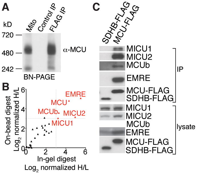
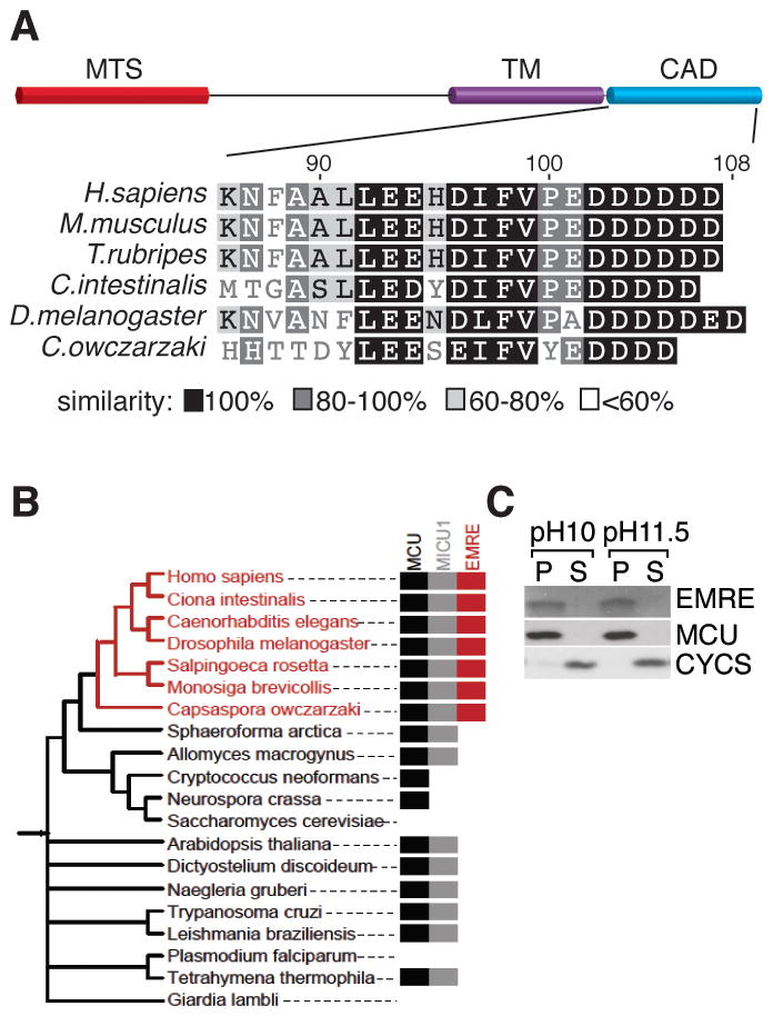
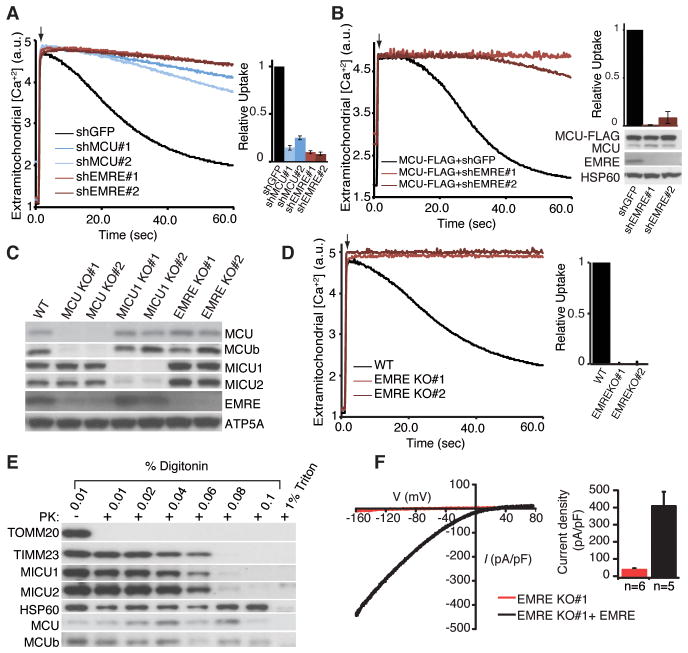
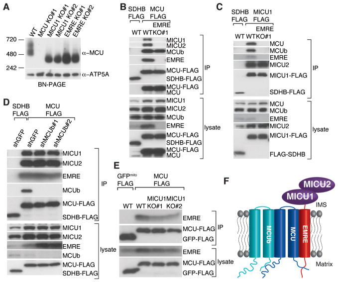
The mitochondrial uniporter is a highly selective calcium channel in the organelle's inner membrane. Its molecular components include the EF-hand-containing calcium-binding proteins mitochondrial calcium uptake 1 (MICU1) and MICU2 and the pore-forming subunit mitochondrial calcium uniporter (MCU). We sought to achieve a full molecular characterization of the uniporter holocomplex (uniplex). Quantitative mass spectrometry of affinity-purified uniplex recovered MICU1 and MICU2, MCU and its paralog MCUb, and essential MCU regulator (EMRE), a previously uncharacterized protein. EMRE is a 10-kilodalton, metazoan-specific protein with a single transmembrane domain. In its absence, uniporter channel activity was lost despite intact MCU expression and oligomerization. EMRE was required for the interaction of MCU with MICU1 and MICU2. Hence, EMRE is essential for in vivo uniporter current and additionally bridges the calcium-sensing role of MICU1 and MICU2 with the calcium-conducting role of MCU.


Figures

Fig. 1
Fig. 1. Affinity purification and proteomic analysis of the uniporter complex, uniplex
(A) MCU-FLAG or control SDHB-FLAG was stably expressed in HEK-293T cells. Proteins from digitonin-permeabilized mitochondria from MCU-FLAG expressing cells and FLAG immunoprecipitations from SDHB-FLAG and MCU-FLAG expressing cells were subjected to Blue Native-PAGE (BN-PAGE) and immunoblotted with MCU antibody. (B) Identification of proteins that interact with MCU-FLAG. MCU-FLAG expressing cells were grown in the presence of heavy amino acids, control HEK-293T cells were grown in the presence of light amino acids. FLAG immunoprecipitates from both samples were mixed and analyzed by mass spectrometry. The ratios of heavy and light proteins annotated with mitochondrial localization from two replicates are shown. Proteins that show significant enrichment in heavy samples are shown in red. (C) Interaction of MCUb, MICU1, MICU2 and EMRE with MCU. MCU-FLAG or control SDHB-FLAG was immunoprecipitated from HEK-293T cells. Immunoprecipitates and cell lysates were analyzed by immunoblotting for the indicated proteins.
Fig. 2
Fig. 2. Domain architecture, phylogeny and membrane association of EMRE
(A) Schematics showing predicted mitochondrial targeting sequence (MTS), transmembrane (TM) and conserved carboxy terminal acidic domain (CAD) of EMRE. The CAD of EMRE from six species was aligned using BLOSUM similarity matrix. (B) EMRE is metazoan-specific. Presence of homologs of MCU/MCUb, MICU1/MICU2 and EMRE across 20 selected species spanning the NCBI taxonomy tree, indicating that EMRE is a metazoan innovation. (C) EMRE is a membrane protein. HEK-293T cell mitochondria were isolated and proteins were extracted with 0.1M Na2CO3 at pH 10 and pH 11.5. EMRE is observed in the insoluble pellet (P) similar to MCU. Cytochrome c (CYCS) is loosely associated with the inner membrane and is detected in the soluble fraction (S).
Fig. 3
Fig. 3. Requirement of EMRE for uniporter activity
(A) EMRE knock down impairs mitochondrial calcium uptake. Digitonin permeabilized HEK-293T cells were incubated with the calcium indicator Oregon Green Bapta 6F (OGB6F). After addition of 50 μM CaCl2 (arrow), the depletion of extracellular calcium due to mitochondrial uptake was monitored by OGB6F fluorescence. Representative traces of mitochondrial calcium uptake from HEK-293T cells with GFP (control), MCU and EMRE knock down are shown. The bar graph shows the rate of calcium uptake relative to shGFP cells (mean ± s.d., n=4). (B) MCU overexpression cannot rescue mitochondrial calcium uptake in EMRE knock down cells. EMRE was knocked down in HEK-293T cells that overexpress MCU-FLAG. The samples were treated as in (A) (n=4). Lysates were analyzed by immunoblotting for indicated proteins. (C) Loss of MCU decreases EMRE and MCUb abundance. MCU, MICU1 and EMRE knockout (KO) HEK-293T cells were generated using TALEN technology, cell lysates from wild type (WT) and two independent knockout clones were prepared and analyzed by immunoblotting for control ATP5A and uniplex proteins (D) Mitochondrial calcium uptake is severely impaired in cells that lack EMRE. Mitochondrial calcium uptake was measured in WT and two independent EMRE KO cell lines as in (A). (E) Loss of EMRE does not change MCU, MCUb, MICU1 and MICU2 submitochondrial localization. Mitochondria were isolated from cells lacking EMRE, incubated with increasing concentrations of digitonin in the presence of proteinase K (PK), samples were analyzed by immunoblotting for outer membrane protein TOMM20, inner membrane protein TIMM23, matrix protein HSP60, as well as uniplex proteins (F) Exemplar trace (red) of a mitoplast derived from EMRE knockout cells demonstrates absent calcium current (IMiCa), whereas typical IMiCa is seen after EMRE is knocked back in (black). Voltage ramps were delivered from −160mV to +80mV for 750ms, using a holding potential of 0mV. (Right) Summary data. Error bars report SEM.
Fig. 4
Fig. 4. Requirement of EMRE for the interaction of MCU with MICU1 and MICU2
(A) Loss of EMRE or MICU1 reduces the size of the uniplex to ~300 kD. Mitochondria from indicated cell lines were isolated, digitonin permeabilized and immunoblotted for MCU after BN-PAGE. EMRE mediates MCU-MICU1 and MICU2 interaction. Stably expressed SDHB-FLAG (control) or MCU-FLAG (B) or MICU1-FLAG (C) was immunoprecipitated from WT or EMRE knockout cells, lysates and immunoprecipitates were analyzed by immunoblotting for indicated proteins. (D) Loss of MCUb does not alter MCU-EMRE, MICU1 and MICU2 interaction. Stably expressed SDHB-FLAG or MCU-FLAG was immunoprecipitated from cells after GFP (control) or MCUb knock down with two different hairpins. Samples were treated and analyzed as in (B). (E) Loss of MICU1 does not alter MCU-EMRE interaction. Plasmid for EMRE expression was co-transfected with plasmids for control mitochondrial GFP-FLAG or MCU-FLAG expression, in WT and MICU1 knockout cells. Samples were treated and analyzed as in (B). (F) Model showing submitochondrial localization and organization of uniplex proteins. MCU is the pore-forming component of the uniplex; MICU1 and MICU2 reside in the IMS where they sense outside calcium and regulate MCU through EMRE. EMRE is a single-pass transmembrane protein. It bridges MCU and MICU1 and MICU2 and is essential for the activity of the uniporter. MCU-MCUb interaction is not mediated by EMRE.

References

    1. Kirichok Y, Krapivinsky G, Clapham DE. The mitochondrial calcium uniporter is a highly selective ion channel. Nature. 2004 Jan 22;427:360. - PubMed
    1. Perocchi F, et al. MICU1 encodes a mitochondrial EF hand protein required for Ca(2+) uptake. Nature. 2010 Sep 16;467:291. - PMC - PubMed
    1. Baughman JM, et al. Integrative genomics identifies MCU as an essential component of the mitochondrial calcium uniporter. Nature. 2011 Aug 18;476:341. - PMC - PubMed
    1. De Stefani D, Raffaello A, Teardo E, Szabo I, Rizzuto R. A forty-kilodalton protein of the inner membrane is the mitochondrial calcium uniporter. Nature. 2011 Aug 18;476:336. - PMC - PubMed
    1. Plovanich M, et al. MICU2, a paralog of MICU1, resides within the mitochondrial uniporter complex to regulate calcium handling. PLoS One. 2013;8:e55785. - PMC - PubMed

Publication types

MeSH terms