Review
. 2014 Feb;37(1):26-36.
doi: 10.1007/s00270-013-0776-y.

Radiological diagnosis and management of epistaxis

Review

Radiological diagnosis and management of epistaxis

Antonín Krajina et al. Cardiovasc Intervent Radiol. 2014 Feb.

Abstract

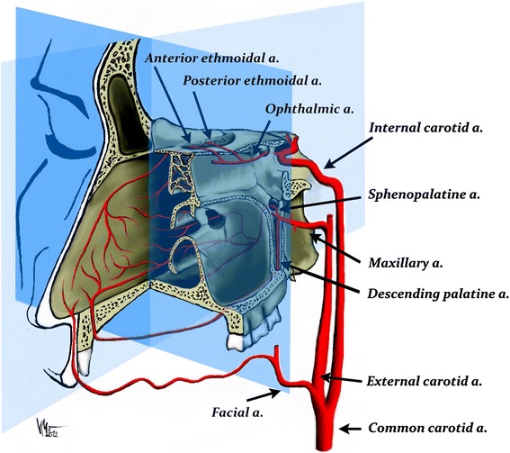
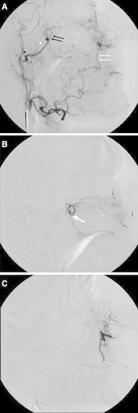
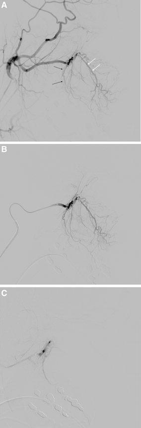
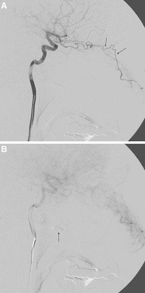
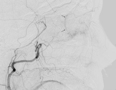
The majority of episodes of spontaneous posterior epistaxis treated with embolisation are idiopathic in nature. The angiographic findings are typically normal. Specific angiographic signs are rare and may include the following: a tumour blush, telangiectasia, aneurysm, and/or extravasation. Selective internal carotid artery (ICA) angiography may show rare causes of epistaxis, such as traumatic or mycotic aneurysms, which require different treatment approaches. Complete bilateral selective external and internal carotid angiograms are essential to evaluation. The images should be analysed for detection of central retinal blush in the external carotid artery (ECA) and anastomoses between the branches of the ECA and ICA. Monocular blindness and stroke are two of the most severe complications. Embolisation aims to decrease flow to the bleeding nasal mucosa while avoiding necrosis of the nasal skin and palate mucosa. Embolisation is routinely performed with a microcatheter positioned in the internal maxillary artery distal to the origin of the meningeal arteries. A guiding catheter should be placed in the proximal portion of the ECA to avoid vasospasm. Embolisation with microparticles is halted when the peripheral branches of the sphenopalatine artery are occluded. The use of coils is not recommended because recurrent epistaxis may occur due to proximal embolization; moreover, the option of repeat distal embolisation is lost. The success rate of embolisation therapy (accounting for late recurrence of bleeding) varies between 71 and 94 %. Results from endoscopic surgery are quite comparable. When epistaxis is refractory to nasal packing or endoscopic surgery, embolisation is the treatment of choice in some centres.


Figures

Fig. 1
Fig. 1
Schematic arterial supply of the sinonasal cavity. The majority of the posterior epistaxis episodes arise from the septum. The arterial branches involved in epistaxis include the internal maxillary artery, the facial artery, and the ophthalmic artery (Courtesy of V. Machova)
Fig. 2
Fig. 2
A External carotid angiogram of a patient with HHT and multiple episodes of severe epistaxis. The target artery for embolisation is the sphenopalatine artery (double black arrows) and the terminal portion of the facial artery (double white arrows). A microcatheter for embolisation should be placed distal to the middle meningeal artery (small black arrowhead) and accessory meningeal artery (small white arrowhead). B Selective internal maxillary angiogram showing the position of a microcatheter (large white arrow). There are separate mucosal hypervascular areas caused by telangiectasias (small black arrows). C Selective facial angiogram that shows the supply to the nasal cavity
Fig. 3
Fig. 3
A Angiographic anatomy of the distal internal maxillary artery. The descending palatine artery (black arrows) outlines the posterior wall and floor of the maxillary antrum. The infraorbital artery (double white arrows) enters the orbit through the infraorbital fissure. B Selective distal internal maxillary artery angiogram in a lateral view. C Completion angiogram after embolisation with microparticles (Courtesy of J. J. Vitek)
Fig. 4
Fig. 4
A Internal carotid angiogram in a patient with recurrent epistaxis. There is a rich collateral supply to the nasal cavity from the ophthalmic artery (arrows). B The cause of such a collateral pathway is previous proximal embolisation of the internal maxillary artery using coils (arrow) (Courtesy of Dr. M. Vavrova)
Fig. 5
Fig. 5
External carotid angiogram showing the supply of the retina (arrows)

References

    1. Melia L, McGarry GW. Epistaxis: update on management. Curr Opin Otolaryngol Head Neck Surg. 2011;19:30–35. doi: 10.1097/MOO.0b013e328341e1e9. - DOI - PubMed
    1. Shukla PA, Chan N, Duffis EJ, Eloy JA, Prestigiacomo CJ, Gandhi CD. Current treatment strategies for epistaxis: a multidisciplinary approach. J Neurointerv Surg. 2012;5(2):1–6. - PubMed
    1. Chiu TW, McGarry GW. Prospective clinical study of bleeding sites in idiopathic adult posterior epistaxis. Otolaryngol Head Neck Surg. 2007;137:390–393. doi: 10.1016/j.otohns.2006.10.035. - DOI - PubMed
    1. Schlosser RJ. Epistaxis. N Engl J Med. 2009;360:784–789. doi: 10.1056/NEJMcp0807078. - DOI - PubMed
    1. Knopfholz J, Lima-Junior E, Précoma-Neto D, Faria-Neto JR. Association between epistaxis and hypertension: a 1 year follow-up after an index episode of nose bleeding in hypertensive patients. Int J Cardiol. 2009;134:e107–e109. doi: 10.1016/j.ijcard.2008.01.018. - DOI - PubMed

LinkOut - more resources