Review
. 2013 Dec;14(12):1062-72.
doi: 10.1038/embor.2013.182. Epub 2013 Nov 15.

Cellular dynamics in the muscle satellite cell niche

Affiliations
Review

Cellular dynamics in the muscle satellite cell niche

C Florian Bentzinger et al. EMBO Rep. 2013 Dec.

Abstract

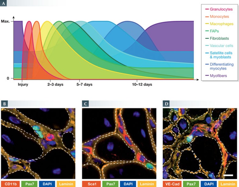
Satellite cells, the quintessential skeletal muscle stem cells, reside in a specialized local environment whose anatomy changes dynamically during tissue regeneration. The plasticity of this niche is attributable to regulation by the stem cells themselves and to a multitude of functionally diverse cell types. In particular, immune cells, fibrogenic cells, vessel-associated cells and committed and differentiated cells of the myogenic lineage have emerged as important constituents of the satellite cell niche. Here, we discuss the cellular dynamics during muscle regeneration and how disease can lead to perturbation of these mechanisms. To define the role of cellular components in the muscle stem cell niche is imperative for the development of cell-based therapies, as well as to better understand the pathobiology of degenerative conditions of the skeletal musculature.


Figures

Figure 1
Figure 1
Overview of tissue histology during mouse skeletal muscle regeneration. (A) A time course of histological changes in regenerating skeletal muscle. H&E staining of uninjured TA muscles and regenerating TA muscles at 5, 10 and 30 days after intramuscular cardiotoxin injection. Regenerating muscles are reduced to mostly mononuclear cells at day 5, but are able to re-establish multinucleated myofibres by day 10. Notably, the nuclei of uninjured myofibres are located at the periphery, whereas those of regenerating muscle fibres are centrally located. Scale bar, 50 μm. (B) Longitudinal view of whole tissue preparations of uninjured (left) and regenerating (right) skeletal muscle. Immunostaining for the extracellular matrix protein laminin (green) labels the basal lamina surrounding myofibres and capillaries. In regenerating conditions, the proliferation of satellite cells can be observed by the increase in the number of Pax7 (red) expressing cells (arrows). DAPI staining of nuclei (blue) reveals accessory cells in the satellite cell niche. Scale bar, 50 μm. H&E, haematoxylin and eosin; TA, tibialis anterior.
Figure 2
Figure 2
Schematic representation of the various cell types involved in muscle regeneration. Within the complexity of regenerating muscles, satellite cells are subject to a distinct environment determined by the spatial and temporal presence of cytokines, growth factors and other cell types.
Figure 3
Figure 3
Participation of non-myogenic cell types in muscle regeneration. (A) The relative presence of immune, fibrotic, vascular and myogenic cell types after muscle injury. (B–D) Immunofluorescence micrographs of tissue sections from regenerating mouse muscles. In their niche, Pax7-positive satellite cells (green) are in close proximity to various non-myogenic cell types (red): (B) CD11b-positive leukocytes; (C) Sca1-positive interstitial cells; and (D) VE-Cad-positive endothelial cells. ECM is shown in orange and nuclei are labelled with DAPI (blue). Scale bar, 10 μm.
Figure 4
Figure 4
Schematic of extrinsic signals in the muscle stem cell niche. Paracrine signals (thin arrows) regulate the recruitment, proliferation rate and differentiation (bold arrows) of each cell type.
None
C Florian Bentzinger, Yu Xin Wang, Nicolas A Dumont & Michael A Rudnicki

Similar articles

Cited by

References

    1. Lieber RL (2010) Skeletal Muscle Structure, Function, and Plasticity. Baltimore, MD, USA: Wolters Kluwer/Lippincott Williams & Wilkins
    1. Mauro A (1961) Satellite cell of skeletal muscle fibers. J Biophys Biochem Cytol 9: 493–495 - PMC - PubMed
    1. Judson RN, Zhang RH, Rossi FM (2013) Tissue-resident mesenchymal stem/progenitor cells in skeletal muscle: collaborators or saboteurs? Febs J 280: 4100–4108 - PMC - PubMed
    1. Pannerec A, Marazzi G, Sassoon D (2012) Stem cells in the hood: the skeletal muscle niche. Trends Mol Med 18: 599–606 - PubMed
    1. Mounier R, Chretien F, Chazaud B (2011) Blood vessels and the satellite cell niche. Curr Top Dev Biol 96: 121–138 - PubMed

Publication types