Review
. 2013 Dec;14(12):1073-83.
doi: 10.1038/embor.2013.181. Epub 2013 Nov 15.

Three wise centromere functions: see no error, hear no break, speak no delay

Affiliations
Review

Three wise centromere functions: see no error, hear no break, speak no delay

Tomoyuki U Tanaka et al. EMBO Rep. 2013 Dec.

Abstract

The main function of the centromere is to promote kinetochore assembly for spindle microtubule attachment. Two additional functions of the centromere, however, are becoming increasingly clear: facilitation of robust sister-chromatid cohesion at pericentromeres and advancement of replication of centromeric regions. The combination of these three centromere functions ensures correct chromosome segregation during mitosis. Here, we review the mechanisms of the kinetochore-microtubule interaction, focusing on sister-kinetochore bi-orientation (or chromosome bi-orientation). We also discuss the biological importance of robust pericentromeric cohesion and early centromere replication, as well as the mechanisms orchestrating these two functions at the microtubule attachment site.


Figures

Figure 1
Figure 1
Three wise centromere functions. (A) The three wise monkeys, which are mystic apes known as Mizaru, Kikazaru and Iwazaru in Japanese. Together they symbolize the proverbial principle to see no evil, hear no evil, speak no evil. (B) The three wise functions of the centromere: orchestration of proper microtubule attachment (see no error), robust sister-chromatid cohesion (hear no break) and early S-phase replication (speak no delay) at the same chromosome site.
Figure 2
Figure 2
Error correction promoted by Aurora B kinase. Aurora B kinase promotes error correction, leading to sister-kinetochore bi-orientation. When tension is not applied on a kinetochore–microtubule attachment, kinetochore phosphorylation by Aurora B causes its turnover (left). According to the Aurora B spatial separation model, on bi-orientation, kinetochores delocalize from Aurora B, which causes kinetochore dephosphorylation and stops the turnover (right). On bi-orientation, kinetochore–microtubule attachment could be stabilized also due to its intrinsic properties.
Figure 3
Figure 3
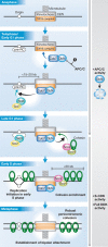
Roles of DDK at kinetochores in budding yeast. DDK (Dbf4–Cdc7) promotes pericentromeric sister-chromatid cohesion and advances the replication of centromeric regions in budding yeast [57]. DDK is recruited to kinetochores during telophase to early G1 phase by the Ctf19 kinetochore complex. The DDK at kinetochores in turn recruits Sld3–Sld7 replication initiation proteins to pericentromeric replication origins in telophase to early G1 phase, as well as the Scc2–Scc4 cohesin loader to centromeres in the late G1 phase. pre-RC, pre-replicative complex.
Figure 4
Figure 4
Cohesin distribution at pericentromeres in budding yeast. DDK at kinetochores enhances the amount of cohesins at centromeres and at pericentromeric regions (up to 20 kb from centromeres) in budding yeast [57]. The graph shows the amount of Scc1 along the indicated chromosome region around the centromere of chromosome XII (CEN12). The Scc1 amount was measured by chromatin immunoprecipitation, followed by high-throughput DNA sequencing (ChIP-seq) [149,150], in DBF4+ (no tag; black line) and DBF4-myc (in which the amount of DDK is reduced at kinetochores; red line) cells. Adapted from Natsume T et al (2013) Mol Cell 50: 661–674 [57].
Figure 5
Figure 5
Roles of DDK at fission yeast pericentromeric heterochromatin. In fission yeast, the heterochromatin protein Swi6 recruits DDK (called Dfp1–Hsk1) to pericentromeres [79]. Dfp1–Hsk1 in turn promotes the recruitment of cohesins and the replication initiation protein Sld3 to pericentromeric regions, leading to robust sister-chromatid cohesion and early S-phase replication of these regions [79,135]. The table on the right shows orthologues in fission and budding yeast.
Figure 6
Figure 6
Profile of replication timing of a budding yeast chromosome. DDK at kinetochores advances replication timing of centromeric regions in budding yeast [57]. The graph shows the profile of replication timing of chromosome XVI. The profile was obtained from high-throughput DNA sequence reads [129] in S phase of wild-type (black dots and line), Dbf4-myc (blue) and ctf19Δ (red) cells. In Dbf4-myc and ctf19Δ cells, the amount of DDK at kinetochores is reduced. Adapted from Natsume T et al (2013) Mol Cell 50: 661–674 [57].
None
Tomoyuki U Tanaka
None
Lesley Clayton
None
Toyoaki Natsume

Similar articles

Cited by

References

    1. Tanaka TU (2002) Bi-orienting chromosomes on the mitotic spindle. Curr Opin Cell Biol 14: 365–371 - PubMed
    1. Gartenberg M (2009) Heterochromatin and the cohesion of sister chromatids. Chromosome Res 17: 229–238 - PubMed
    1. Tanaka TU (2010) Kinetochore-microtubule interactions: steps towards bi-orientation. EMBO J 29: 4070–4082 - PMC - PubMed
    1. Kitamura E, Tanaka K, Kitamura Y, Tanaka TU (2007) Kinetochore microtubule interaction during S phase in Saccharomyces cerevisiae. Genes Dev 21: 3319–3330 - PMC - PubMed
    1. Hegemann JH, Fleig UN (1993) The centromere of budding yeast. Bioessays 15: 451–460 - PubMed

Publication types

Substances

LinkOut - more resources