Review
. 2013 Dec;14(12):1050-61.
doi: 10.1038/embor.2013.173. Epub 2013 Nov 15.

Building and remodelling Cullin-RING E3 ubiquitin ligases

Affiliations
Review

Building and remodelling Cullin-RING E3 ubiquitin ligases

John R Lydeard et al. EMBO Rep. 2013 Dec.

Abstract

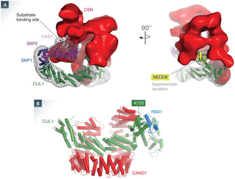
Cullin-RING E3 ubiquitin ligases (CRLs) control a plethora of biological pathways through targeted ubiquitylation of signalling proteins. These modular assemblies use substrate receptor modules to recruit specific targets. Recent efforts have focused on understanding the mechanisms that control the activity state of CRLs through dynamic alterations in CRL architecture. Central to these processes are cycles of cullin neddylation and deneddylation, as well as exchange of substrate receptor modules to re-sculpt the CRL landscape, thereby responding to the cellular requirements to turn over distinct proteins in different contexts. This review is focused on how CRLs are dynamically controlled with an emphasis on how cullin neddylation cycles are integrated with receptor exchange.


Conflict of interest statement

J.W.H. is a consultant for Millenium Pharmaceutics.

Figures

Figure 1
Figure 1
Architecture of human cullin–RING E3 ubiquitin ligase system. The number of human SRs for each CRL complex is indicated on the left. CUL4A and CUL4B are represented as a single CRL. The CRL regulatory apparatus is composed of the neddylation system, the deneddylation system and the SR-exchange factor. UBC12 is the neddylation E2 for RBX1-based CRL complexes, whereas UBE2F is the E2 for CRL5–RBX2. DCN1 is a co-E3 for both RBX1 and RBX2. The CSN deneddylates CRLs. CAND1 is a CRL exchange factor that interacts with both the amino- and carboxy-terminal regions of the cullin–RING complex. CSN subunits are indicated, an asterisk indicates that CSN5 is the catalytic subunit. CRL, cullin–RING E3 ubiquitin ligase; CSN, COP9 signalosome; N, NEDD8; S, substrate; SR, substrate receptor; U, ubiquitin.
Figure 2
Figure 2
Structure of selected cullin-binding components of neddylation machinery and the effect of cullin neddylation on SCF structure. (A) DCN1 (violet) co-recruits UBC12's N-terminal helix (cyan) and the WHB subdomain at the carboxy-terminal from a cullin (CUL1, green) [39]. (B) Close-up view of panel A, with DCN1 shown in surface view coloured by electrostatic potential (red, negative; blue, positive), which highlights the hydrophobic pocket that binds to UBC12's N-acetyl methionine. (C) Model for an unneddylated but fully assembled CRL in complex with an E2–ubiquitin intermediate, based on superposition of structures of CUL1–RBX1–SKP1–SKP2F-box, SKP1–SKP2–CKS1–p27 phosphopeptide and RING–UBCH5–E2 [17,125,126,127]. The p27 phosphopeptide is shown in spheres. The substrate is distal from the E2–ubiquitin active site. (D) Model of a neddylated CRL showing the potential for RBX1 RING domain rotation, which is based on superimposing common features of CUL1–RBX1 with NEDD8–CUL5CTD–RBX1 [61]. NEDD8 is shown in yellow covalently linked to the repositioned portion of CUL1. The location of the RBX1 RING domain found in some unneddylated CRL structures is shown in blue, with alternative positions found in neddylated structures shown in sky and cyan. CRL, cullin–RING E3 ubiquitin ligase.
Figure 3
Figure 3
Structures of the CSN–SCF and CAND1–CUL1–RBX1 complexes. (A) Two views of the structure obtained by electron microscopy of catalytically inactive CSN bound to SKP1 (dark blue)–SKP2 (purple)–CKS1 (pink)–NEDD8 (yellow)–CUL1 (green)–RBX1, with density for these proteins shown as grey mesh and for CSN in red. Because NEDD8, the CUL1 WHB domain to which it is linked, and the RBX1 RING domain are similar sizes, only their approximate locations are indicated [41]. (B) Crystal structure of CUL1 (green)—RBX1 (blue)–CAND1 (red). The NEDD8 acceptor lysine (Lys 720) on CUL1 is shown in spheres [66].
Figure 4
Figure 4
CAND1 as a substrate receptor exchange factor. (A) Pathways for activation, SR exchange and deneddylation of CRLs. This model is based on the proposed role for CAND1 as an SR exchange factor [35], the role of the CSN complex as both a catalytic and binding-dependent inhibitor of CRLs, the action of which can be reversed by substrate [36,40,41], and the stoichiometries of the various components in vivo [36]. The model initiates with empty cullin (step 1), formed, for example, by new synthesis. This empty cullin could be engaged by a newly synthesized SR1 module (step 2) or potentially integrated into a CAND1 complex for exchange (not shown for clarity). The basal CRL complex could either enter into a CAND1-dependent exchange cycle (step 3) or could become rapidly neddylated and engage substrates (steps 4 and 5). As substrate abundance is diminished, CRLs can associate with CSN, and become deneddylated (step 6), followed by dissociation of the CRL complex to re-enter the cycle. The CAND1-exchange pathway leads to dissociation of SR1 and association of SR2 (either a pre-existing or newly synthesized module) through a proposed intermediate in which CAND1 and SR2 associate simultaneously with the cullin complex (step 3). Exchange of SR1 for SR2 would change which substrates are targeted in the cell. (B) Structure depicting how the β-hairpin of CAND1 (red) bound to CUL1 (green) clashes with SKP1 (blue), based on superimposing CUL1–RBX1–CAND1 and CUL1–RBX1–SKP1–SKP2F-box structures [17,66]. The arrow denotes the location of the β-hairpin in CAND1. Some residues of SKP1 that are absent in the crystal structure might further clash with CAND1. CRL, cullin–RING E3 ubiquitin ligase; SR, substrate receptor.

References

    1. Mizushima N, Komatsu M (2011) Autophagy: renovation of cells and tissues. Cell 147: 728–741 - PubMed
    1. Hershko A, Ciechanover A (1998) The ubiquitin system. Annu Rev Biochem 67: 425–479 - PubMed
    1. Metzger MB, Pruneda JN, Klevit RE, Weissman AM (2013) RING-type E3 ligases: master manipulators of E2 ubiquitin-conjugating enzymes and ubiquitination. Biochim Biophys Acta [Epub 6 Jun] ; DOI: 10.1016/j.bbamcr.2013.05.026 - DOI - PMC - PubMed
    1. Wenzel DM, Stoll KE, Klevit RE (2011) E2s: structurally economical and functionally replete. Biochem J 433: 31–42 - PMC - PubMed
    1. Komander D, Rape M (2012) The ubiquitin code. Annu Rev Biochem 81: 203–229 - PubMed

Publication types