. 2013 Nov 15:3:3224.
doi: 10.1038/srep03224.

Establishment of a novel lingual organoid culture system: generation of organoids having mature keratinized epithelium from adult epithelial stem cells

Affiliations

Establishment of a novel lingual organoid culture system: generation of organoids having mature keratinized epithelium from adult epithelial stem cells

Hiroko Hisha et al. Sci Rep. .

Abstract

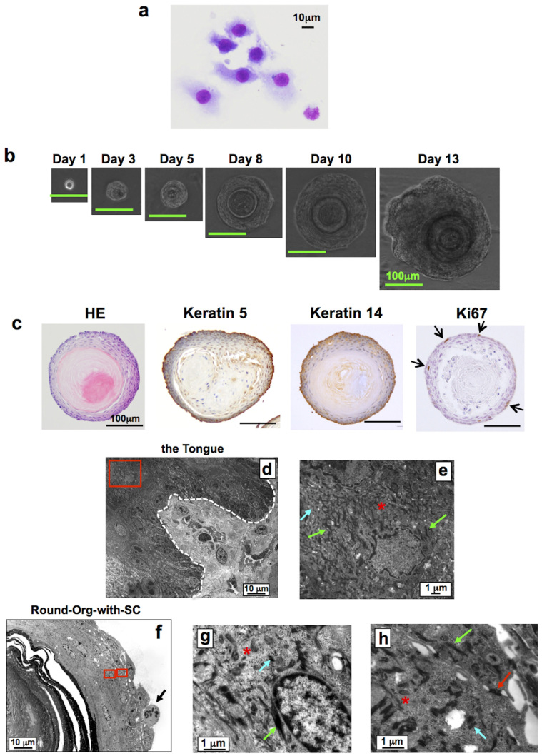
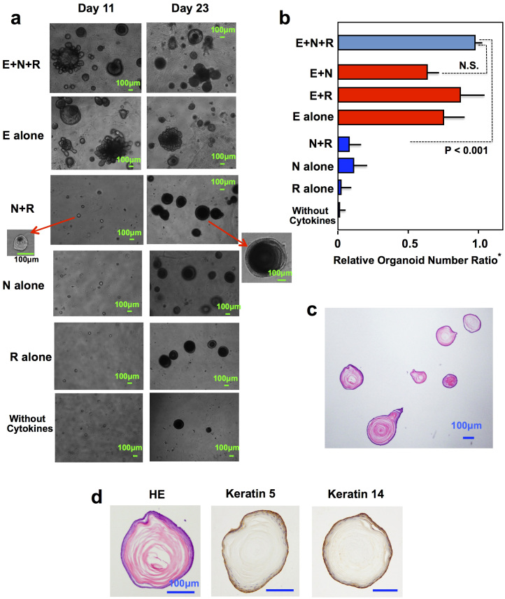
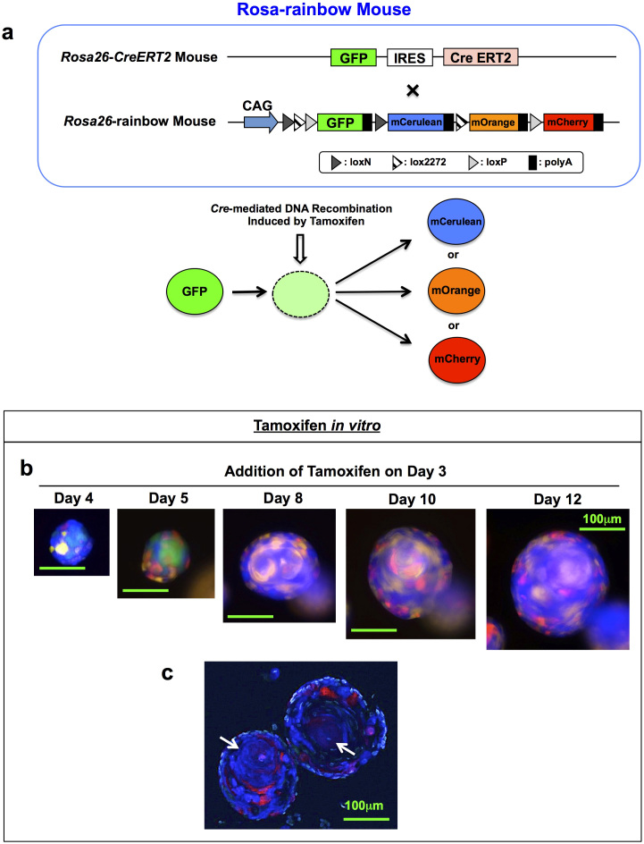
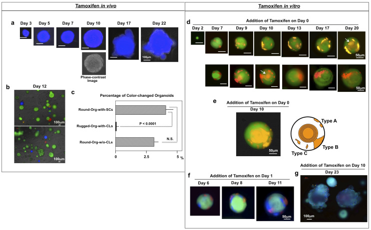
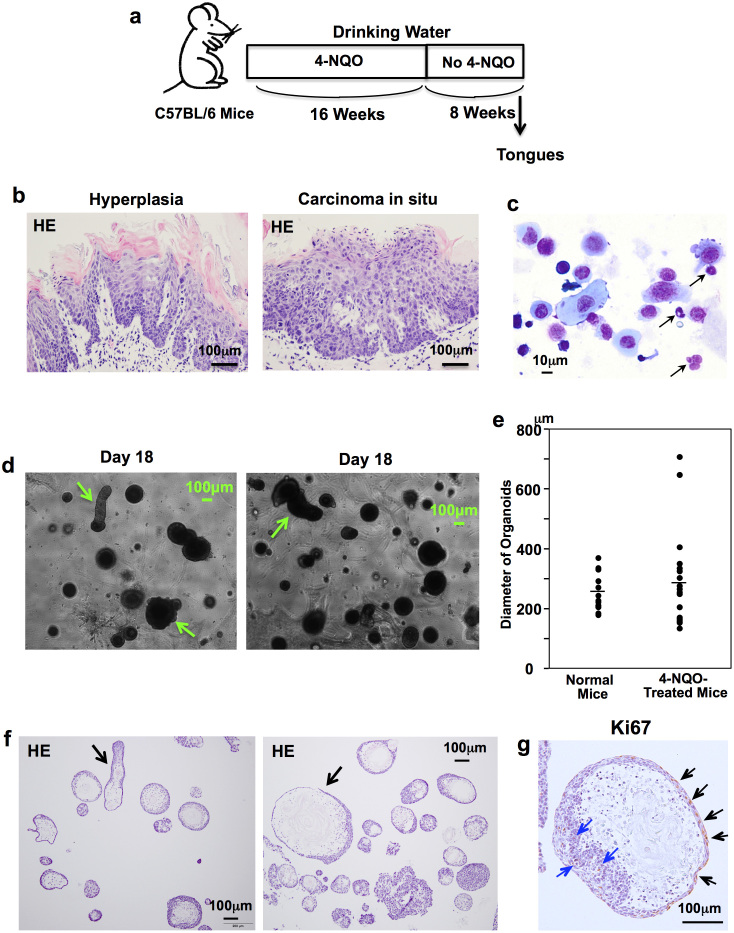
Despite the strong need for the establishment of a lingual epithelial cell culture system, a simple and convenient culture method has not yet been established. Here, we report the establishment of a novel lingual epithelium organoid culture system using a three-dimensional matrix and growth factors. Histological analyses showed that the generated organoids had both a stratified squamous epithelial cell layer and a stratum corneum. Very recently, we showed via a multicolor lineage tracing method that Bmi1-positive stem cells exist at the base of the epithelial basal layer in the interpapillary pit. Using our new culture system, we found that organoids could be generated by single Bmi1-positive stem cells and that in the established organoids, multiple Bmi1-positive stem cells were generated at the outermost layer. Moreover, we observed that organoids harvested at an early point in culture could be engrafted and maturate in the tongue of recipient mice and that the organoids generated from carcinogen-treated mice had an abnormal morphology. Thus, this culture system presents valuable settings for studying not only the regulatory mechanisms of lingual epithelium but also lingual regeneration and carcinogenesis.


Figures

Figure 1
Figure 1. Morphological analysis of round-shape organoids with concentric cell arrangement.
(a) HE staining of separated LECs. (b) Time course of lingual organoid growth. LECs were cultured in the organoid culture system in the presence of EGF + noggin + R-spondin1 and round-shape organoids with concentric cell arrangement were generated. Phase-contrast image. (c) Histological analysis of organoids. Organoids were separated from Matrigel using dispase on day 14 of culture and their paraffin sections were stained with HE reagents. The organoid sections were also stained with anti-keratin 5 and keratin 14 antibodies. Positive cells were observed in the outer periphery of the organoids. Staining for Ki67 showed that some cells actively proliferated in the outer periphery (arrows). (d) Transmission electron micrograph of the normal tongue. The dotted line outlines the border between epithelium and lamina propria mucosa. Basal and intermediate epithelium (upper side of the dotted line) and lamina propria mucosa (lower side of the dotted line). (e) Higher magnification views of the boxed areas in (d). Many keratohyaline granules (blue arrow), keratin fibers (green arrow) and some lamellar granules (indicated by asterisk) were detected in the cytoplasm. (f) Transmission electron micrograph of organoids containing stratum corneum. Stratum corneum in the center and flattened cells surrounding the stratum corneum were observed. In the outer side of the organoid, a cell having aggregated chromosome (indicated by arrow) was detected. (g,h) Higher magnification views of the boxed areas in (f). Many keratohyaline granules (blue arrow), keratin fibers (green arrow) and lamellar granules (indicated by asterisk) were detected in the cytoplasm. Moreover, formation of desmosome between cells was observed (red arrow).
Figure 2
Figure 2. Characterization of organoids generated in different cytokine combinations.
(a) Skewed generation into the round-shape organoids in the cultures without EGF and in the culture without cytokine. LECs were cultured in the organoid culture system in the presence of various cytokine combinations or in the absence of cytokines. In the cultures with the cytokine combinations which did not contain EGF and in the culture without cytokines, organoids were very small (less than 50 μm) on day 11 of culture, but thereafter only round-shape organoids were generated. Phase-contrast image. (b) Comparison of organoid number in different cytokine combinations. The number of organoids was counted on day 23 of culture. (3 wells/sample). Mean ± SD of 3 independent experiments. *: Relative organoid number ratio = No. of organoids in culture of cytokine combination other than EGF + noggin + R-spondin1 or in culture without cytokines/No. of organoids in culture of the cytokine combination of EGF + noggin + R-spondin1. N.S.: not significant. E: EGF, N: noggin and R: R-spondin1. (c) HE staining of organoids obtained from the noggin + R-spondin1 combination. The formed organoids were separated from Matrigel on day 23 of culture and their frozen sections were stained with HE reagents. The organoids were composed of very thick stratum corneum and very thin epithelial layer. (d) Keratin 5 and keratin 14 staining of organoids obtained from the noggin + R-spondin1 combination. The cells in the outer periphery of the organoids were stained positively with anti-keratin 5 and 14 antibodies.
Figure 3
Figure 3. Observation of organoid-forming process using color-labeled cells.
(a) Schematic representations of gene constructs and Cre-mediated fluorescent color change in Rosa-rainbow mice. In Rosa-rainbow mice, fluorescent colors of individual cells change from green to different color (red-, orange- or blue-color) by Cre-mediated excision of floxed cassettes induced by tamoxifen and their descendant cells retain the changed color. (b) A representative growth pattern of organoid. LECs of Rosa-rainbow mice were cultured in the organoid culture system in the presence of EGF + noggin + R-spondin1 for 3 days and active form of tamoxifen was added to the culture. Individual cells were labeled by red-, orange- or blue-color after the Cre induction and the organoids showed mosaic patterns composed of the colored cells on day 5 of culture. Over time, the mosaic patterns disappeared and domains showing blue color expanded gradually and most parts of the organoids were occupied with blue color. Fluorescence image. (c) Frozen section of organoids collected on day 14 of culture. Most parts of the organoids were occupied with blur-colored cells. Stratum corneum is indicated by arrows. Nuclei were stained with Hoechst 33342. Fluorescence image.
Figure 4
Figure 4. Capacity of Bmi1-positive cells to generate organoids.
(a–c) in vivo administration of tamoxifen. LECs obtained from Bmi1-rainbow mice that had been injected with tamoxifen 2 days before were cultured in the organoid culture system in the presence of EGF + noggin + R-spondin1. (a) Time course of organoid growth in a blue-colored round-Org-with-SC. Overlay of fluorescence image and phase-contrast image. (b) Representative photographs of organoids on day 12 of culture. Most organoids showed green color, but some round-shape organoids showed red-, orange- or blue-color. Overlay of fluorescence image and phase-contrast image. (c) Percentage of color-changed organoids in each type of organoids. The total number of generated organoids and the number of color-changed organoids were counted on day 12. N.S.: not significant. (d–g) in vitro administration of tamoxifen. Tamoxifen was added to the organoid culture medium of Bmi1-rainbow mice at the beginning of culture (d,e), on day 1 (f) or on day 10 of culture (g). Fluorescence image. (d) Time course of organoid growth in a round-Org-with-SC containing orange-colored or red-colored cells derived from Bmi1-positive cells. Fluorescence image indicates that these organoids had colored stratum corneum (indicated by arrows). (e) A representative round-Org-with-SC on day 10 of culture and a schematic representation of three different types of growth patterns of Bmi1-positive cells. (f) Time course of organoid growth in a round-Org-with-SC having two or more-colored cells. Addition of tamoxifen on day 1 of culture induced red-, orange- or blue-colored cells in a single organoid. (g) A representative photograph of round-Org-with-SC on day 23 of culture. Many colored cells were detected in each organoid, when tamoxifen was added on day 10.
Figure 5
Figure 5. Expansion of engrafted organoids in the tongues of recipient mice.
(a) Three-day-organoids generated from LECs of Bmi1-rainbow mice. Phase-contrast image shows that the organoids are 20–30 μm in diameter and do not have stratum corneum showing that these organoids are immature. (b) Experimental protocol of organoid transplantation experiments. (c) A representative frozen section of the recipient tongue collected at 1 week after engrafting. Enlarged organoids were detected in the muscle layer. Stratum corneum is indicated by arrows. Nuclei were stained with Hoechst 33342. Fluorescence image. (d) A representative frozen section of the recipient tongue collected at 2 weeks after engrafting. Nuclei were stained with Hoechst 33342. Fluorescence image. (e) HE staining of paraffin-embedded sections of recipient tongues at 2 weeks after grafting.
Figure 6
Figure 6. Lingual organoids generated from LECs of carcinogen-treated mice.
(a) Experimental protocol of 4-NQO administration. (b) HE staining of paraffin-embedded tongue sections obtained from carcinogen-treated mice. (c) HE staining of separated LECs. (d) Overview of generated organoids on day 18 of culture. Very large or slender organoids (indicated by arrows) were detected. Phase-contrast image. (e) Comparison of organoid diameters between normal mice and carcinogen-treated mice. There was no significant difference in organoid diameters between normal mice and carcinogen-treated mice. (f) HE staining of frozen sections of organoids. Organoids were separated from Matrigel using dispase on day 18 of culture and their frozen sections were stained with HE reagents. Abnormal shapes of organoids (indicated by arrow) were observed. (g) Ki67 staining of organoids. Frozen sections of the organoids were stained with anti-Ki67 antibody. Ki67-positive cells were detected in the middle epithelial layer (indicated by blue arrows) and in the outer side of the organoids (indicated by black arrows).

Similar articles

Cited by

References

    1. Reiner D. J. et al. Genetic analysis of tongue size and taste papillae number and size in recombinant inbred strains of mice. Chem Senses 33, 693–707 (2008). - PMC - PubMed
    1. Mbiene J.-P., Maccallum D. K. & Mistretta C. M. Organ cultures of embryonic rat tongue support tongue and gustatory papilla morphogenesis in vitro without intact sensory ganglia. J Comp Neurol 377, 324–340 (1997). - PubMed
    1. Zhou Y., Liu H.-X. & Mistretta C. M. Bone morphogenic proteins and noggin: Inhibiting and inducing fungiform taste papilla development. Develop Biol 297, 198–213 (2006). - PubMed
    1. Ookura T. et al. Fibroblast and epidermal growth factors modulate proliferation and neural cell adhesion molecule expression in epithelial cells derived from the adult mouse tongue. In Vitro Cell Dev Biol Anim 38, 365–372 (2002). - PubMed
    1. Luo X., Okubo T., Randell S. & Hogan B. L. M. Culture of endodermal stem/progenitor cells of the mouse tongue. In Vitro Cell Dev Biol Anim 45, 44–54 (2009). - PubMed

Publication types

MeSH terms

Substances