. 2013 Nov 15;342(6160):825-9.
doi: 10.1126/science.1244373.

Iron(IV)hydroxide pK(a) and the role of thiolate ligation in C-H bond activation by cytochrome P450

Affiliations

Iron(IV)hydroxide pK(a) and the role of thiolate ligation in C-H bond activation by cytochrome P450

Timothy H Yosca et al. Science. .

Abstract

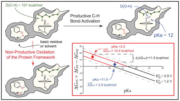
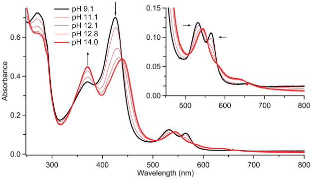
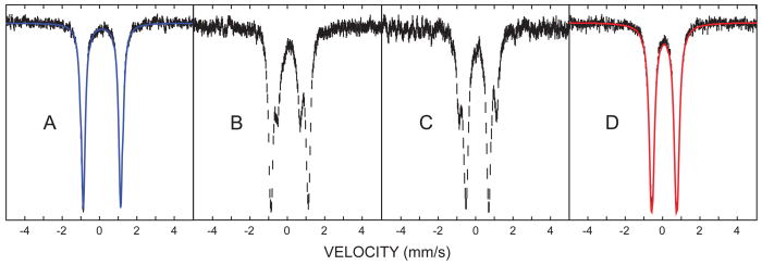
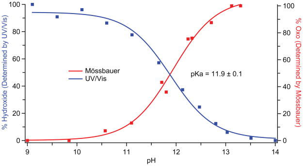
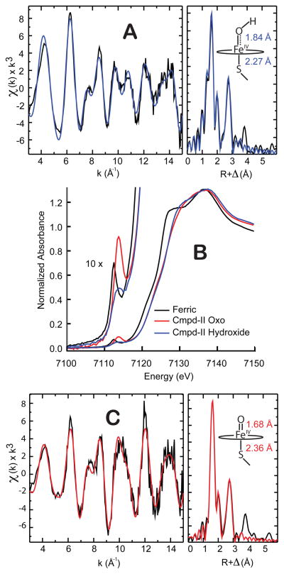
Cytochrome P450 enzymes activate oxygen at heme iron centers to oxidize relatively inert substrate carbon-hydrogen bonds. Cysteine thiolate coordination to iron is posited to increase the pK(a) (where K(a) is the acid dissociation constant) of compound II, an iron(IV)hydroxide complex, correspondingly lowering the one-electron reduction potential of compound I, the active catalytic intermediate, and decreasing the driving force for deleterious auto-oxidation of tyrosine and tryptophan residues in the enzyme's framework. Here, we report on the preparation of an iron(IV)hydroxide complex in a P450 enzyme (CYP158) in ≥90% yield. Using rapid mixing technologies in conjunction with Mössbauer, ultraviolet/visible, and x-ray absorption spectroscopies, we determine a pK(a) value for this compound of 11.9. Marcus theory analysis indicates that this elevated pK(a) results in a >10,000-fold reduction in the rate constant for oxidations of the protein framework, making these processes noncompetitive with substrate oxidation.


Figures

Fig. 1
Fig. 1
Productive and non-productive pathways for compound I decay. During productive C-H bond activation, compound I (a ferryl-radical species) abstracts hydrogen from substrate to yield compound II (an iron(IV)hydroxide complex) and a substrate radical. The non-productive pathway shown involves the oxidation of a tyrosine residue contained within the protein framework, with the phenolic proton being transferred to B:, a basic residue or solvent molecule, upon tyrosine oxidation. The inset shows the relative free energies (ΔGrel, Eq. 5) for the productive and non-productive pathways as a function of the compound II pKa for the range of aqueous tyrosine potentials. When ΔGrel is negative, the productive pathway is thermodynamically preferred. As a result of the ± 2 kcal/mol error in Eq. 2, a small range of tyrosine potentials could yield a given ΔGrel. The uncertainty in the solid lines as a result of this error is ± 0.1 V.
Fig. 2
Fig. 2
UV/Visible spectra of CYP158-II at varying pH. Samples were prepared in a double-mix stopped-flow experiment. 30μM ferric CYP158 (10 mM Tris-HCl, pH 9.0) was first mixed with 150 μM m-CPBA and held until maximum formation of CYP158-II was achieved (2.5 seconds). This solution was then mixed with a strongly buffered solution (containing 200 mM phosphate, 200 mM carbonate, pH adjusted with KOH) to obtain the desired final pH. Mixing was 1:1:1. The inset shows a magnification of the 450–800 nm region.
Fig. 3
Fig. 3
Mössbauer spectra of CYP158-II at varying pH. (A) pH 9.0 (B) pH 11.8 (C) 12.2 (D) 13.3. Samples were prepared in a double-mix freeze-quench experiment. 6 mM protein was mixed 2:1 with 60 mM m-CPBA to form CYP158-II at pH 9. This species was then mixed 3:1 with an arginine/NaOH buffer (pH 14). The strength of the arginine/NaOH buffer was varied (10mM to 108.16mM) to achieve the desired final pH. The reaction mixture was sprayed into liquid ethane 7 ms after the first mix (SM Materials and Methods). Fits for the intermediate pH samples (B and C) are shown in fig. S2.
Fig. 4
Fig. 4
pH titration curves for CYP158-II. Data obtained from stopped-flow UV/Visible (blue) and Mössbauer spectroscopies (red) yield the same pKa value of 11.9. UV/Visible data points were obtained from the change in absorbance at 423 nm (Fig. 2). Mössbauer data points were obtained from the ratio of the areas of the iron(IV)oxo and iron(IV)hydroxide subspectra (fig S2).
Fig. 5
Fig. 5
X-ray absorption spectroscopy. A) EXAFS of CYP158-II at pH 9.0. B) X-ray absorption edges. C) EXAFS of CYP158-II at pH 13.3. All EXAFS samples were analyzed by Mössbauer spectroscopy prior to data collection. See tables S1 and S2 for further details.

Comment in

References

    1. Schlichting I, et al. Science. 2000;287:1615. - PubMed
    1. Rittle J, Green MT. Science. 2010;330:933. - PubMed
    1. Sligar SG. Science. 2010;330:924. - PMC - PubMed
    1. Bollinger JM, Jr, Broderick JB. Curr Opin Chem Biol. 2009;13:51. - PubMed
    1. Groves JT. Proc Natl Acad Sci USA. 2003;100:3569. - PMC - PubMed

Publication types