Review
. 2014 Feb;29(2):263-73.
doi: 10.1093/ndt/gft443. Epub 2013 Nov 13.

The iron cycle in chronic kidney disease (CKD): from genetics and experimental models to CKD patients

Affiliations
Review

The iron cycle in chronic kidney disease (CKD): from genetics and experimental models to CKD patients

Kimberly Zumbrennen-Bullough et al. Nephrol Dial Transplant. 2014 Feb.

Abstract

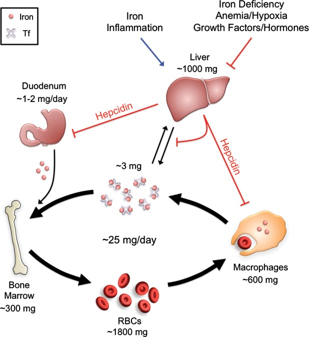
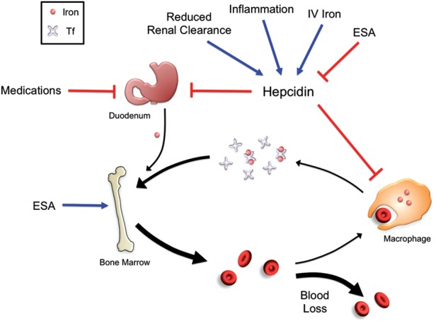
Iron is essential for most living organisms but iron excess can be toxic. Cellular and systemic iron balance is therefore tightly controlled. Iron homeostasis is dysregulated in chronic kidney disease (CKD) and contributes to the anemia that is prevalent in this patient population. Iron supplementation is one cornerstone of anemia management in CKD patients, but has not been rigorously studied in large prospective randomized controlled trials. This review highlights important advances from genetic studies and animal models that have provided key insights into the molecular mechanisms governing iron homeostasis and its disturbance in CKD, and summarizes how these findings may yield advances in the care of this patient population.

Keywords: anemia; chronic kidney disease; hepcidin; iron; review.


Figures

FIGURE 1:
FIGURE 1:
Systemic iron regulation. Iron is absorbed by the duodenum where it is released into the circulation via the iron exporter ferroportin to be loaded onto transferrin (Tf). The majority of iron is utilized by red blood cells (RBCs) for the synthesis of the hemoglobin, requiring ∼25 mg of iron per day. The daily requirements for intestinal iron uptake are only 1–2 mg per day due to efficient recycling of iron from RBCs. Iron recycling is performed primarily by reticuloendothelial macrophages which phagocytize senescent RBCs and then export iron via ferroportin back into the circulating pool of Tf-bound iron. Excess iron is also stored within hepatocytes. Hepcidin regulates systemic iron balance by inducing ferroportin degradation to inhibit iron absorption from the duodenum and iron release from macrophage and hepatocyte stores. Hepcidin production in the liver is stimulated by iron and inflammation to limit iron availability, while hepcidin production is inhibited by iron deficiency, anemia and hypoxia to increase iron availability. Several other growth factors and steroid hormones have recently been demonstrated to suppress hepcidin expression in the liver, including EGF, HGF, testosterone and estrogen.
FIGURE 2:
FIGURE 2:
Enterocyte iron uptake. Dietary iron absorption occurs via the reduction of ferric (Fe3+) iron to ferrous (Fe2+) iron by ferrireductases such as DCYTB. Ferrous iron is then transported across the apical membrane of duodenal enterocytes by the symporter DMT1. Heme is also an important source of dietary iron, although the mechanism for heme uptake is unclear. Heme oxygenase 1 (HO1) is thought to facilitate the degradation of heme into iron, biliverdin and carbon monoxide. Cytoplasmic iron can be stored by the ferritin complex, utilized by various molecular enzymes or exported into the bloodstream by ferroportin (FPN). The multicopper ferroxidase hephaestin (HEPH) works in conjunction with ferroportin to facilitate iron export coupled with oxidization of Fe2+ to Fe3+ and loading onto Tf.
FIGURE 3:
FIGURE 3:
Molecular regulation of hepcidin by iron and inflammation. Increased systemic iron stimulates the production of the ligand bone morphogenetic protein 6 (BMP6), which binds to the BMP Type I (ALK2/ALK3) and II (ACTRIIA) receptors, and the co-receptor HJV to stimulate phosphorylation of the SMAD1/5/8 intracellular signaling molecules. Phosphorylated SMAD 1/5/8 binds to SMAD4 and translocates to the nuclease to activate hepcidin transcription. The mechanism by which the hemochromatosis protein HFE and/or TFR2 regulate hepcidin expression is unknown but appears to involve an interaction with the BMP-SMAD signaling pathway. It has been proposed that an interaction between HFE and TFR1 is reduced under high iron conditions due to competitive binding of holotransferrin to TFR1. Displaced HFE could then associate with TFR2 and possibly the HJV-BMP receptor complex to regulate hepcidin. Inflammation also stimulates hepcidin production, in part via a canonical janus kinase (JAK)/signal transducer and activator of transcription (STAT) pathway in which inflammation increases interleukin 6 (IL6) binding to the IL6-receptor (IL6R) and thereby stimulating phosphorylation of JAKs and STAT3. Phosphorylated-STAT3 homodimers translocate to the nuclease and bind to the hepcidin promoter to stimulate hepcidin expression. Other mediators of inflammation and infection can also regulate hepcidin expression in this context (not shown). A mechanism of crosstalk between inflammatory signals and BMP signaling has been proposed in which inflammation induces activin B, which binds to BMP receptors to stimulate SMAD1/5/8 phosphorylation. SMADs and STAT3 may also interact at the level of the hepcidin promoter.
FIGURE 4:
FIGURE 4:
Disordered iron balance in CKD. Chronic inflammation and reduced renal clearance in patients with CKD lead to increased levels of hepcidin, which reduces duodenal iron uptake and iron release from cellular iron stores. Intestinal iron uptake is also inhibited by medications such as phosphate binders and antacids. ESAs stimulate increased iron usage for erythropoiesis, while blood loss due to frequent phlebotomy, blood trapping in the dialysis apparatus and gastrointestinal bleeding further deplete the circulating iron pool. Iron administration stimulates hepcidin expression, which can paradoxically worsen the iron restriction, while ESAs have an inhibitory effect on hepcidin expression.

References

    1. Hentze MW, Muckenthaler MU, Andrews NC. Balancing acts: molecular control of mammalian iron metabolism. Cell. 2004;117:285–297. - PubMed
    1. Kovac S, Anderson GJ, Baldwin GS. Gastrins, iron homeostasis and colorectal cancer. Biochim Biophys Acta. 2011;1813:889–895. - PMC - PubMed
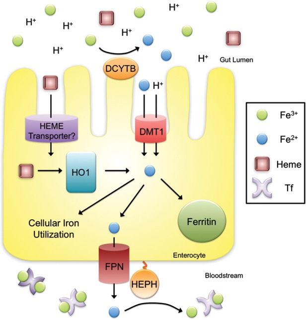
    1. McKie AT, Barrow D, Latunde-Dada GO, et al. An iron-regulated ferric reductase associated with the absorption of dietary iron. Science. 2001;291:1755–1759. - PubMed
    1. Gunshin H, Starr CN, Direnzo C, et al. Cybrd1(duodenal cytochrome b) is not necessary for dietary iron absorption in mice. Blood. 2005;16:16. - PMC - PubMed
    1. Fleming MD, Trenor CC, III, Su MA, et al. Microcytic anaemia mice have a mutation in Nramp2, a candidate iron transporter gene. Nat Genet. 1997;16:383–386. - PubMed

Publication types