. 2014 Jan 10;289(2):868-84.
doi: 10.1074/jbc.M113.501064. Epub 2013 Nov 14.

Automethylation activities within the mixed lineage leukemia-1 (MLL1) core complex reveal evidence supporting a "two-active site" model for multiple histone H3 lysine 4 methylation

Affiliations

Automethylation activities within the mixed lineage leukemia-1 (MLL1) core complex reveal evidence supporting a "two-active site" model for multiple histone H3 lysine 4 methylation

Anamika Patel et al. J Biol Chem. .

Abstract

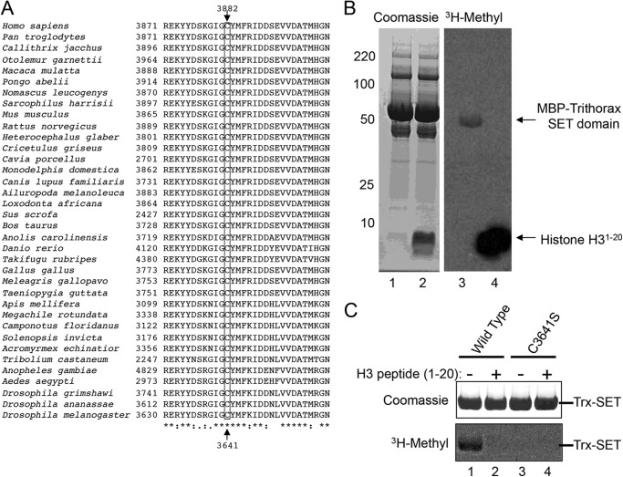
The mixed lineage leukemia-1 (MLL1) core complex predominantly catalyzes mono- and dimethylation of histone H3 at lysine 4 (H3K4) and is frequently altered in aggressive acute leukemias. The molecular mechanisms that account for conversion of mono- to dimethyl H3K4 (H3K4me1,2) are not well understood. In this investigation, we report that the suppressor of variegation, enhancer of zeste, trithorax (SET) domains from human MLL1 and Drosophila Trithorax undergo robust intramolecular automethylation reactions at an evolutionarily conserved cysteine residue in the active site, which is inhibited by unmodified histone H3. The location of the automethylation in the SET-I subdomain indicates that the MLL1 SET domain possesses significantly more conformational plasticity in solution than suggested by its crystal structure. We also report that MLL1 methylates Ash2L in the absence of histone H3, but only when assembled within a complex with WDR5 and RbBP5, suggesting a restraint for the architectural arrangement of subunits within the complex. Using MLL1 and Ash2L automethylation reactions as probes for histone binding, we observed that both automethylation reactions are significantly inhibited by stoichiometric amounts of unmethylated histone H3, but not by histones previously mono-, di-, or trimethylated at H3K4. These results suggest that the H3K4me1 intermediate does not significantly bind to the MLL1 SET domain during the dimethylation reaction. Consistent with this hypothesis, we demonstrate that the MLL1 core complex assembled with a catalytically inactive SET domain variant preferentially catalyzes H3K4 dimethylation using the H3K4me1 substrate. Taken together, these results are consistent with a "two-active site" model for multiple H3K4 methylation by the MLL1 core complex.

Keywords: Automethylation; Chromatin Histone Modification; Enzyme Mechanisms; Epigenetics; Histone Methylation; Leukemia; Self-methylation.


Figures

FIGURE 1.
FIGURE 1.
MLL1 SET domain undergoes an automethylation reaction that is inhibited by histone H3. A, surface representation of the nucleosome core particle (PDB code 1KX5 (61)) is shown with the position of H3K4 indicated. The dashed box encompasses residues 1–20 of histone H3. The box below shows a schematic of histone H3 peptides used in this investigation, consisting of amino acid residues 1–20. The positions of lysine or trimethyl-lysine residues are indicated. B, comparison of enzymatic activity of the MLL1 SET domain (amino acid residues 3811–3969, MLL3811) among histone H3 peptides that were unmodified (H3Unmodified, lanes 1 and 2) or previously trimethylated at Lys-4 (H3K4me3, lane 3), or trimethylated at Lys-9 (H3K9me3, lane 4). C, MLL1 automethylation is abolished when Asn-3906 is replaced with alanine.
FIGURE 2.
FIGURE 2.
MLL1 automethylation is irreversible and relatively robust. A, MLL3745 was incubated with 1 μm [3H]AdoMet for 2 h, followed by the addition of 1 mm unlabeled (cold) AdoMet or an equivalent volume of reaction buffer. Reactions were quenched at the indicated time points with 1× SDS loading buffer and separated by SDS-PAGE. Upper panel shows Coomassie Blue-stained gel, and lower panel shows the fluorogram of the same gel. B, 5 μm MLL3745 was incubated with 25 μm [3H]AdoMet in the presence or absence of 250 μm histone H3 peptide (residues 1–20). Reactions were quenched at the indicated time points with 1× SDS loading buffer and separated by SDS-PAGE. Histone peptide and MLL3811 bands at the different time points were excised from the gels and quantitated by liquid scintillation counting. Open circles show the rate of histone H3 peptide methylation, and closed circles show the rate of MLL3811 automethylation in the absence of histone H3 peptide. Linear regressions gave slopes of 704 ± 46 (R2 = 0.98) and 212 ± 12 (R2 = 0.99) for histone H3 and MLL3811 automethylation reactions, respectively. C, apparent Km determination for AdoMet in the histone H3 methylation reaction catalyzed by MLL1. The change in counts from LSC (Δcpm, h−1) versus AdoMet concentration was fit with nonlinear least squares regression to the Michaelis-Menten equation with an apparent Km value for AdoMet of 10.4 ± 3.1. D, apparent Km determination for AdoMet in the MLL1 automethylation reaction. Changes in relative intensity per h (Δ%I, h−1) were plotted against AdoMet concentration (0–20 μm) and fit with the Michaelis-Menten equation with an apparent Km value of 6.5 ± 1.5. Inset shows automethylation activity over the concentration range 0–50 μm, which displays a pattern of substrate inhibition at 50 μm AdoMet.
FIGURE 3.
FIGURE 3.
MLL1 automethylates in an intramolecular fashion. A, wild type MLL3811 was incubated with the larger catalytically inactive N3906A MLL3745 variant and 1 μm [3H]AdoMet in the absence (lanes 1 and 3) or presence of histone H3 peptide (lanes 2 and 4). Reactions were quenched after 4 h and separated by SDS-PAGE. Left panel shows Coomassie Blue-stained gel, and right panel shows the fluorogram of the same gel. B, same as in A except that wild type MLL3745 was incubated with the catalytically inactive N3906A MLL3811 variant in the absence (lanes 1 and 3) and presence of histone H3 peptide (lanes 2 and 4). C, log-log plot showing the concentration dependence of MLL1 automethylation velocity. The data were fit with linear regression showing a slope of 0.7 (R2 = 0.99). Error bars represent one standard deviation from the mean from two independent experiments.
FIGURE 4.
FIGURE 4.
Mass spectrometry identifies Cys-3882 as a site for MLL1 automethylation. A, sequence of the MLL3745 construct; B, table of predicted trypsin proteolytic peptides and expected masses. C, MALDI-TOF spectrum of the trypsinized MLL3745 protein after preincubation with 1 mm AdoMet. The peptide at m/z 960.4 is indicated and is 14 Da larger than the expected GIGCYMFR peptide at 946.4. D, ESI LC MS/MS spectrum of the doubly charged precursor at m/z = 480.8, which corresponds to the methylated peptide GIGCYMFR. The table below displays all predicted and observed b- and y-ions for the unmodified and modified forms of the peptide, respectively. Each of the peptides highlighted in yellow are 14 Da greater than predicted, indicating that Cys-3882 is methylated. E, MALDI-TOF spectrum of the trypsinized C3882S MLL3745 protein. The peak corresponding to the GIGSYFMR peptide is indicated at m/z 930.5. F, comparison of histone methylation and automethylation activities of wild type and C3882A/S MLL3745 proteins in the presence and absence of histone H3 peptide. Reactions were quenched at 8 h and separated by 4–12% BisTris PAGE and visualized by Coomassie Blue staining (upper panel) and fluorography (lower panel).
FIGURE 5.
FIGURE 5.
Cysteine 3882 is located in the SET-I subdomain of MLL1. A, schematic representation of the C-terminal SET domain and Win motif of MLL1. The position of Cys-3882 in the SET-I subdomain is indicated. The SET-I subdomain separates the more widely conserved SET-N and SET-C subdomains. B, surface representation of the three-dimensional structure of the MLL1 SET domain (PDB code 2W5Z (12)). Histone H3 peptide is indicated with carbon atoms in white, nitrogen atoms in blue, and oxygen atoms in red. The co-factor product AdoHcy is indicated with carbon atoms in green, nitrogen atoms in blue, oxygen atoms in red, and sulfur atoms in yellow. The position of cysteine 3882 is indicated and highlighted in yellow. C, blow-up of the dashed box in B shows that the Cys-3882 side chain sulfur is greater than 10 Å away from the predicted position of the AdoMet sulfonium moiety. D, superposition of Cα atoms of the SET domains from MLL1 (magenta, PDB code 2W5Z (12)) and Dim5 (green, PDB code 1PEG (30)). The positions of Cys-3882 in MLL1 and the equivalent residue in Dim5 (Val-203) are indicated.
FIGURE 6.
FIGURE 6.
SET domain automethylation is conserved throughout metazoan evolution. A, ClustalW2 multiple sequence alignment of SET-I subdomain amino acids among metazoan MLL1 orthologs. The position of Cys-3882 in MLL1 (Homo sapiens numbering) is boxed and indicated at the top. The equivalent position (Cys-3641) in the D. melanogaster Trithorax protein is indicated at the bottom. B, SET domain from the D. melanogaster Trithorax protein was purified as an MBP fusion and incubated with [3H]AdoMet in the presence (lanes 2 and 4) and absence (lanes 1 and 3) of histone H3 peptide (residues 1–20). The Coomassie Blue-stained gel is shown on the left and fluorogram on the right. C, comparison of automethylation activities of wild type and C3641S Trithorax SET domains in the presence and absence of histone H3 peptide.
FIGURE 7.
FIGURE 7.
MLL1 automethylation is reduced when assembled into the MLL1 core complex. A, automethylation of MLL3745 in the absence (lanes 1 and 2) or presence (lanes 3 and 4) of the WDR5-RbBP5-Ash2L subcomplex. Enzymes (7 μm) were incubated with 1 μm [3H]AdoMet in the presence or absence of 300 μm histone H3 peptide for 8 h. Quenched samples were separated by 4–12% PAGE and visualized by Coomassie Blue staining (upper panel) and fluorography (lower panel). B, comparison of automethylation activities of MWRAD complexes assembled with a full-length (residues 1–538) or truncated form of RbBP5 (residues 1–402). Assays were carried out as described above. The positions of Ash2L and RbBP5 are indicated with the boxes. C, Ash2L is not methylated in the context of WRAD in the absence of MLL1. Assays were visualized by fluorography as described above. D, MLL1 does not methylate Ash2L in the absence of WDR5 and RbBP5. E, Ash2L methylation depends on the catalytic activity of the MLL1 SET domain. Ash2L methylation was compared between MWRAD complexes assembled with wild type (lanes 1 and 3) or N3906A (lanes 2 and 4) MLL3745 SET domains in the absence of histone H3. Lanes 1 and 2 are Coomassie Blue-stained gel, and lanes 3 and 4 are the fluorogram of the same gel. F, Ash2L methylation depends on the interaction of MLL1 with WRAD. Wild type (lanes 5–8) and R3765A (lanes 1–4) variants of MLL3745 were assayed in the presence and absence of a stoichiometric amount of WRAD and 300 μm histone H3 peptide for 8 h as described above. G, log-log plot showing the concentration dependence of Ash2L methylation within the context of the MLL1 core complex (MWRAD). The data were fit with linear regression showing a slope of 0.99 ± 0.1 (R2 = 0.93). Error bars represent one standard deviation from the mean from two independent experiments.
FIGURE 8.
FIGURE 8.
MLL1 and Ash2L automethylation reactions distinguish one- versus two-active site models for multiple H3K4 methylation. A, comparison of MLL1 and Ash2L automethylation activities in the absence (lane 1) or presence (lanes 2–5) of full-length recombinant histone H3 proteins that were unmodified or previously mono-, di-, or trimethylated at lysine 4 as MLA (from Active Motif). The upper panel shows the Coomassie Blue-stained gel, and the lower panel is the fluorogram of the same gel. B, ImageJ densitometry of the fluorogram in A. The vertical axis shows percent intensity of each band relative to the intensity of Ash2L or MLL1 methylation in Fig. 8A, lane 1. Histone H3 bands were saturated at this exposure and were not included in the densitometry. C, comparison of MLL1 and Ash2L automethylation activities in the presence or absence of histone H3 peptides (residues 1–21, purchased from Millipore) that were unmodified or previously mono-, di-, or trimethylated at lysine 4. The left-most panel shows the Coomassie Blue-stained gel; the middle panel shows fluorogram after an overnight exposure (18 h), and the right panel shows the fluorogram of the same gel after a 5-day exposure. On the right is ImageJ densitometry of the fluorograms showing amounts of Ash2L or MLL1 methylation relative to their respective intensities in the absence of histone peptide (1st lane).
FIGURE 9.
FIGURE 9.
WRAD preferentially utilizes the H3K4me1 substrate when assembled with the catalytically inactive N3906A MLL1 variant. The MLL1(N3906A) variant was assembled with WRAD, and the complex was purified by size-exclusion chromatography (Superdex 200, GE Healthcare). Complex was assayed with different concentrations of [3H]AdoMet (0–500 μm) with a fixed concentration of unmodified histone H3 peptide (500 μm) (residues 1–20, H3K4me0) or 500 μm H3K4me1 peptide. Samples were quenched at various time points with 1× SDS loading buffer and separated by 4–12% SDS-PAGE. Histone H3 peptide bands were excised from the gel and quantitated by liquid scintillation counting as described under “Experimental Procedures.” Error bars represent the standard error of measurement from two independent experiments. The unpaired Student's t test in Prism (GraphPad) was used to test for statistical significance. p values < 0.05 are indicated.
FIGURE 10.
FIGURE 10.
Model for multiple lysine methylation by MLL1 core complex. A, MLL1 SET domain (M) is shown in pink with the SET-I lobe and Win motif indicated. B, MLL1 undergoes automethylation at Cys-3882 in the presence of AdoMet and absence of histone H3. C, WRAD binding to the Win motif of MLL1 positions N-terminal sequences of Ash2L (A) near the SET domain of MLL1. In the absence of histone H3, Ash2L can then be methylated by MLL1. D, nucleosome (yellow/green) binding to the MLL1 core complex positions the histone H3 N-terminal tail in the MLL1 SET domain active site where it undergoes monomethylation at lysine 4 (H3K4me1). E, H3K4me1 intermediate diffuses away from the MLL1 SET domain, rendering it accessible for automethylation or Ash2L methylation activities. F, H3K4me1 intermediate undergoes dimethylation at active site 2, which is composed of sequences from WRAD and a surface from MLL1 that is distinct from the canonical SET domain active site. G, the MLL1 core complex dissociates from the nucleosome.

Similar articles

Cited by

References

    1. Djabali M., Selleri L., Parry P., Bower M., Young B. D., Evans G. A. (1992) A trithorax-like gene is interrupted by chromosome 11q23 translocations in acute leukaemias. Nat. Genet. 2, 113–118 - PubMed
    1. Gu Y., Nakamura T., Alder H., Prasad R., Canaani O., Cimino G., Croce C. M., Canaani E. (1992) The t(4;11) chromosome translocation of human acute leukemias fuses the ALL-1 gene, related to Drosophila trithorax, to the AF-4 gene. Cell 71, 701–708 - PubMed
    1. Tkachuk D. C., Kohler S., Cleary M. L. (1992) Involvement of a homolog of Drosophila trithorax by 11q23 chromosomal translocations in acute leukemias. Cell 71, 691–700 - PubMed
    1. Ziemin-van der Poel S., McCabe N. R., Gill H. J., Espinosa R., 3rd, Patel Y., Harden A., Rubinelli P., Smith S. D., LeBeau M. M., Rowley J. D., et al. (1991) Identification of a gene, MLL, that spans the breakpoint in 11q23 translocations associated with human leukemias. Proc. Natl. Acad. Sci. U.S.A. 88, 10735–10739 - PMC - PubMed
    1. Patel A., Dharmarajan V., Vought V. E., Cosgrove M. S. (2009) On the mechanism of multiple lysine methylation by the human mixed lineage leukemia protein-1 (MLL1) core complex. J. Biol. Chem. 284, 24242–24256 - PMC - PubMed

Publication types

MeSH terms

LinkOut - more resources