Clinical Trial
. 2014 Jan 3;289(1):520-8.
doi: 10.1074/jbc.M113.511147. Epub 2013 Nov 14.

Tubulation of endosomal structures in human dendritic cells by Toll-like receptor ligation and lymphocyte contact accompanies antigen cross-presentation

Affiliations
Clinical Trial

Tubulation of endosomal structures in human dendritic cells by Toll-like receptor ligation and lymphocyte contact accompanies antigen cross-presentation

Ewoud B Compeer et al. J Biol Chem. .

Abstract

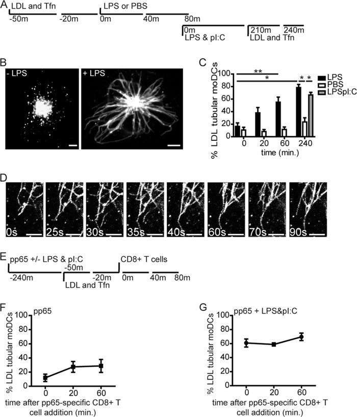
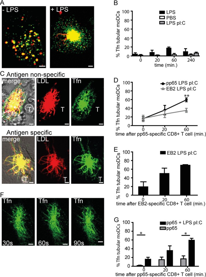
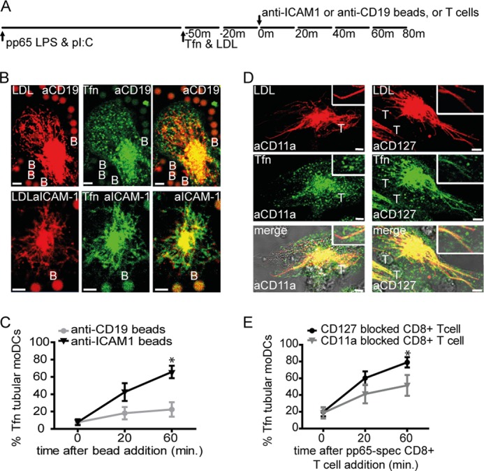
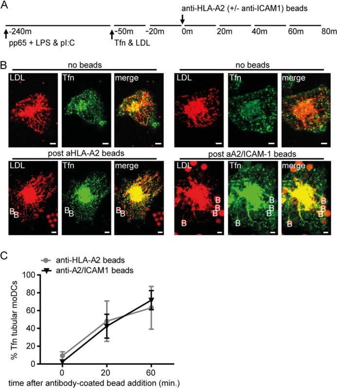
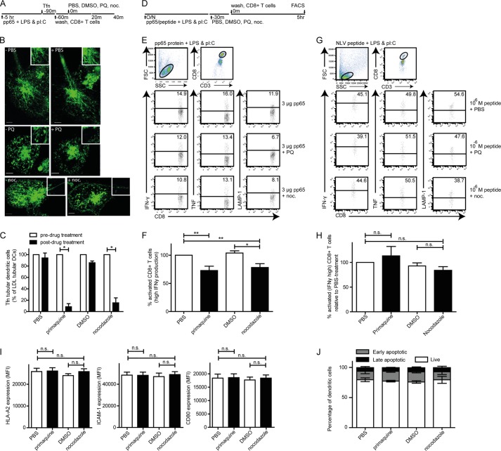
Mouse dendritic cells (DCs) can rapidly extend their Class II MHC-positive late endosomal compartments into tubular structures, induced by Toll-like receptor (TLR) triggering. Within antigen-presenting DCs, tubular endosomes polarize toward antigen-specific CD4(+) T cells, which are considered beneficial for their activation. Here we describe that also in human DCs, TLR triggering induces tubular late endosomes, labeled by fluorescent LDL. TLR triggering was insufficient for induced tubulation of transferrin-positive endosomal recycling compartments (ERCs) in human monocyte-derived DCs. We studied endosomal remodeling in human DCs in co-cultures of DCs with CD8(+) T cells. Tubulation of ERCs within human DCs requires antigen-specific CD8(+) T cell interaction. Tubular remodeling of endosomes occurs within 30 min of T cell contact and involves ligation of HLA-A2 and ICAM-1 by T cell-expressed T cell receptor and LFA-1, respectively. Disintegration of microtubules or inhibition of endosomal recycling abolished tubular ERCs, which coincided with reduced antigen-dependent CD8(+) T cell activation. Based on these data, we propose that remodeling of transferrin-positive ERCs in human DCs involves both innate and T cell-derived signals.

Keywords: Cell-Cell Interaction; Cross-presentation; Dendritic Cells; Endosomes; Intracellular Trafficking; Live Cell Confocal Microscopy; Recycling; T Cell Receptor; Toll-like Receptor (TLR); Tubulation.


Figures

FIGURE 1.
FIGURE 1.
TLR stimulation of human dendritic cells triggers remodeling of late endosomes into tubular structures. A, schematic outline of live cell confocal microscopy experiment in B and C. Fluorescent cargo LDL-DiI and Tfn-Alexa Fluor 647 were stained after 30-min incubation the late and recycling endosomes, respectively, just before or after stimulation with LPS or PBS. B, confocal image of moDCs with vesicular (left) or tubular (right) LDL+ endosomes (60 min 37 °C, 200 ng/ml LPS). C, percentage of moDCs expressing tubular LDL+ endosomes. Time points indicate a 20-min time window immediately before treatment and around indicated time points (20, 60 min); PBS (white bars), 200 ng/ml LPS (black bars), or mix of 200 ng/ml LPS and 5 μg/ml poly(I:C) (gray bars). Data represent mean ± S.E. (error bars) of four independent experiments. D, time-lapse captures of tubular LDL+ endosomes in LPS-treated moDCs. Time points indicate seconds. E, schematic outline of live cell confocal microscopy experiment in F and G. F and G, percentage of moDCs expressing tubular LDL+ endosomes after culture in the presence of pp65 (3 μg of antigen, 4-h time point) and pp65-specific CD8+ T cells (1:1 ratio) in the absence (F) or presence of LPS and poly(I:C) combined (G). Data represent mean ± S.E. of at least three independent experiments using two-tailed Mann-Whitney U test. *, p < 0.05; **, p < 0.01. Scale bars, 5 μm.
FIGURE 2.
FIGURE 2.
Efficient recycling endosome tubular remodeling in human dendritic cells requires cognate T cell interaction. A, representative images of moDCs with LDL+ late endosomes (red) and Tfn+ recycling endosomes (green) in the absence or presence of LPS stimulation (60 min, 200 ng/ml). B, percentage of LDL+ tubular moDCs expressing tubular Tfn+ endosomes prior to stimulation (t = 0) or approximately at the indicated time points. Black bars, 200 ng/ml LPS; gray bars, mix of 200 ng/ml LPS and 5 μg/ml poly(I:C). Data represent mean ± S.E. (error bars) of at least three independent experiments. C, representative images of moDCs upon CD8+ T cell contact in antigen-independent (upper images) or antigen-dependent manner (lower images). Red, LDL; green, Tfn; yellow, co-localization of LDL and Tfn; T, pp65-specific CD8+ T cell. D, percentage of LDL+ tubular moDCs expressing tubular Tfn+ endosomes after 4-h culture in the presence of 3 μg HCMV-derived pp65/LPS/poly(I:C) (black circles) or EBV-derived EB2-LPS-poly(I:C) (gray triangles) at the indicated time points. Two-tailed, Mann-Whitney U test p < 0.01 was used. Data represent mean ± S.E. of at least four independent experiments. E, percentage of LDL+ tubular moDCs expressing tubular Tfn+ endosomes after 4-h culture in the presence of 3 μg of EBV-derived EB2 with LPS and poly(I:C) (black circles) at the indicated time points. Data represent mean ± S.E. of at least three independent experiments. F, time-lapse captures of Tfn+ tubular endosomes in moDCs (time points indicate seconds). Scale bars, 5 μm. G, percentage of LDL+ tubular moDCs expressing tubular Tfn+ endosomes upon culture in the presence of 3 μg of HCMV antigen pp65 (4 h) in the absence (gray bars) or presence of LPS-poly(I:C) (black bars). Data represent mean ± S.E. of at least three independent experiments; *, p < 0.05.
FIGURE 3.
FIGURE 3.
ICAM-1 clustering provokes tubulation of Tfn+ endosomal recycling compartments in human dendritic cells. A, schematic outline of live cell confocal microscopy experiment in B and D. B, representative images of moDCs upon anti-CD19 (upper images) or anti-ICAM-1 (lower images) mAb-coated beads contact. C, percentage of LDL+ tubular moDCs expressing tubular Tfn+ endosomes after 4 h culture in the presence of 3 μg of HCMV-derived pp65-LPS-poly(I:C), prior to stimulation (t = 0) or at the indicated time points upon the addition of anti-CD19 (gray circles), anti-ICAM-1 (black triangles) mAb-coated beads (1:4 DC/bead ratio). One-tailed, Mann-Whitney U test was used. *, p < 0.05. Data represent mean ± S.E. (error bars) of four independent experiments. D, representative images of moDCs upon CD11a (LFA-1, left three images) or CD127 (IL7R, right three images) blocking pp65-specific CD8+ T cell contact. E, percentage of LDL+ tubular moDCs expressing tubular Tfn+ endosomes after 4 h culture in the presence of 3 μg of HCMV-derived pp65/LPS/poly(I:C), prior to stimulation (t = 0) or at the indicated time points upon the addition of CD11a (LFA-1, gray triangles) or CD127 (IL7R, black circles) blocking pp65-specific CD8+ T cells (1:1 DC:T cell ratio). One-tailed, Mann-Whitney U test was used. *, p < 0.05. Data represent mean ± S.E. of six independent experiments. Boxes are enlarged part of images. Red, LDL; green, Tfn; yellow, co-localization of LDL and Tfn; T, CD11a or CD127-blocked pp65-specific CD8+ T cell; B, mAb-coated bead. Scale bars, 5 μm.
FIGURE 4.
FIGURE 4.
Class I MHC clustering or simultaneous ICAM-1-Class I MHC clustering, provokes tubulation of Tfn+ endosomal recycling compartments in human dendritic cells. A, schematic outline of live cell confocal microscopy experiment in B. B, representative images of moDCs upon anti-HLA-A2 mAb (left six images) or anti-ICAM-1/anti-HLA-A2 (right six images). Red, LDL; green, Tfn; yellow, co-localization of LDL and Tfn; B, mAb-coated beads. Scale bars, 5 μm. C, percentage of LDL+ tubular moDCs expressing tubular Tfn+ endosomes after 4-h culture in the presence of 3 μg of HCMV-derived pp65-LPS-poly(I:C) prior to stimulation (t = 0) or at the indicated time points upon the addition of anti-ICAM-1/anti-HLA-A2 (black triangles) or anti-HLA-A2 mAb (gray circles)-coated beads (1:4 DC:bead ratio). One-tailed, Mann-Whitney U test was used. *, p < 0.05. Data represent mean ± S.E. (error bars) of three independent experiments.
FIGURE 5.
FIGURE 5.
Disintegration of tubular Tfn+ endosomal recycling compartments in human dendritic cells associates with reduced ability of dendritic cells to activate antigen-specific CD8+ T cells. A, schematic outline of live cell confocal microscopy experiment in B and C. B and C, representative images (B) and the percentage (C) of selected 3-μg HCMV-derived pp65-LPS-poly(I:C)-laden moDCs after 1 h of co-culture with pp65-specific CD8+ T cells and pretreatment (left three images in B, white bars in C) or after 30 min of the indicated drug treatment (right three images in B, black bars in C). PQ, primaquine, 50 μm; noc, nocodazole, 10 μm; green, Tfn. Scale bar, 5 μm. Data represent the mean ± S.E. (error bars) of at least four independent experiments. D, schematic outline of experimental setup for E–H. E, human moDCs were loaded (overnight, 37 °C) with 0 or 3 μg of HCMV-derived pp65 in the presence of 200 ng/ml LPS and 5 μg/ml poly(I:C). MoDCs were treated (30 min, 37 °C) with either 50 μm primaquine biphosphate, 1 μg/ml nocodazole, or carrier controls PBS, and DMSO, followed by extensive washing (three washes). Next, pp65-specific CD8+ T cells were added for co-culture with the treated moDCs (1:1, 5 h, 37 °C). DC-mediated activation of antigen-specific CD8+ T cells was measured by analysis of induced production of IFNγ, TNF, or surface-expressed LAMP-1 high (bar in dot plot is background, based on 1% cytokine-positive CD8+ T cells upon culture with LPS and poly(I:C)-treated moDCs in absence of antigen). F, percentage of antigen-specific activated CD8+ T cells, determined relative to matched PBS treated-moDCs. G, human moDCs were loaded (overnight, 37 °C) with pp65-derived NLV-peptide in the presence of 200 ng/ml LPS and 5 μg/ml poly(I:C). MoDCs were treated (30 min, 37 °C) with either 50 μm primaquine biphosphate, 1 μg/ml nocodazole, or carrier controls PBS, and DMSO, followed by extensive washing (three washes). Next, pp65-specific CD8+ T cells were added for co-culture with the treated moDCs (1:1, 5 h, 37 °C) and antigen-specific CD8+ T cell activation was determined. H, percentage of antigen-specific activated CD8+ T cells, determined relative to matched PBS-treated moDCs. Human moDCs were loaded (overnight, 37 °C) with 3 μg of HCMV-derived pp65 in the presence of 200 ng/ml LPS and 5 μg/ml poly(I:C). MoDCs were treated (30 min, 37 °C) with either 50 μm primaquine biphosphate, 1 μg/ml nocodazole, or carrier controls PBS, and DMSO, followed by extensive washing (three washes). Thereafter, HLA-A2, ICAM-1, and CD80 expression on (I) or viability (J) of moDCs was determined by flow cytometry analysis. Data represent mean ± S.E. (error bars) of at least three independent experiments. Two-tailed, Mann-Whitney U test was used. *, p < 0.05; **, p < 0.01.

Similar articles

Cited by

References

    1. Grant B. D., Donaldson J. G. (2009) Pathways and mechanisms of endocytic recycling. Nat. Rev. Mol. Cell Biol. 10, 597–608 - PMC - PubMed
    1. Sönnichsen B., De Renzis S., Nielsen E., Rietdorf J., Zerial M. (2000) Distinct membrane domains on endosomes in the recycling pathway visualized by multicolor imaging of Rab4, Rab5, and Rab11. J. Cell Biol. 149, 901–914 - PMC - PubMed
    1. Compeer E. B., Flinsenberg T. W., van der Grein S. G., Boes M. (2012) Antigen processing and remodeling of the endosomal pathway: requirements for antigen cross-presentation. Front. Immunol. 3, 37. - PMC - PubMed
    1. Villadangos J. A., Schnorrer P. (2007) Intrinsic and cooperative antigen-presenting functions of dendritic cell subsets in vivo. Nat. Rev. Immunol. 7, 543–555 - PubMed
    1. Burgdorf S., Schölz C., Kautz A., Tampé R., Kurts C. (2008) Spatial and mechanistic separation of cross-presentation and endogenous antigen presentation. Nat. Immunol. 9, 558–566 - PubMed

Publication types

MeSH terms

Substances