. 2014 Feb 11;129(6):660-72.
doi: 10.1161/CIRCULATIONAHA.113.005746. Epub 2013 Nov 14.

Survivin-induced abnormal ploidy contributes to cystic kidney and aneurysm formation

Affiliations

Survivin-induced abnormal ploidy contributes to cystic kidney and aneurysm formation

Wissam A Aboualaiwi et al. Circulation. .

Abstract

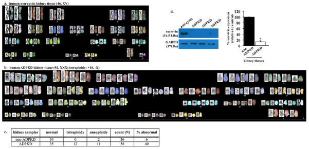
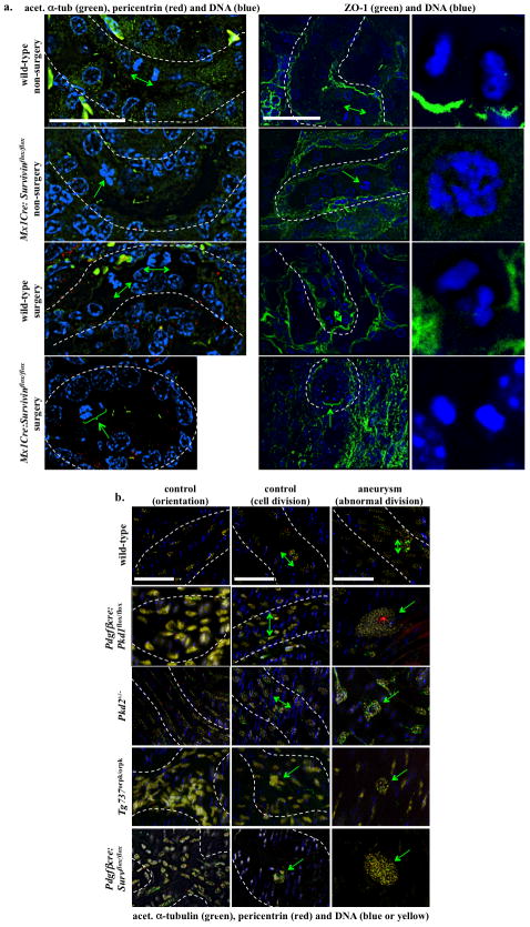
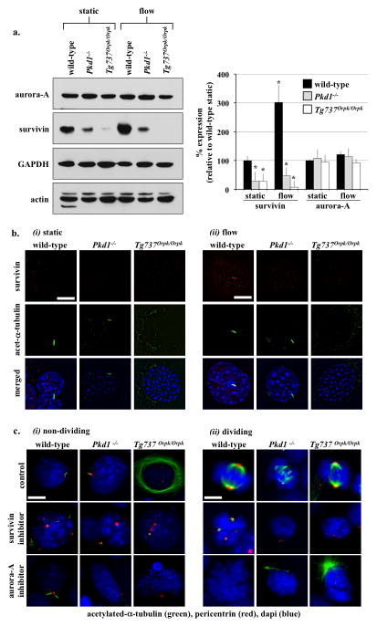
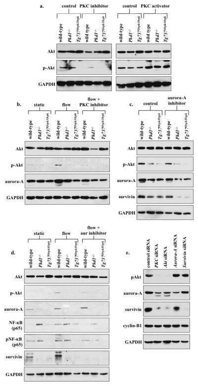
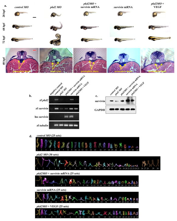
Background: Cystic kidneys and vascular aneurysms are clinical manifestations seen in patients with polycystic kidney disease, a cilia-associated pathology (ciliopathy). Survivin overexpression is associated with cancer, but the clinical pathology associated with survivin downregulation or knockout has never been studied before. The present studies aim to examine whether and how cilia function (Pkd1 or Pkd2) and structure (Tg737) play a role in cystic kidney and aneurysm through survivin downregulation.

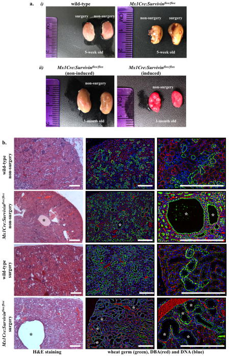
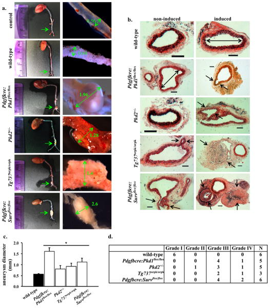
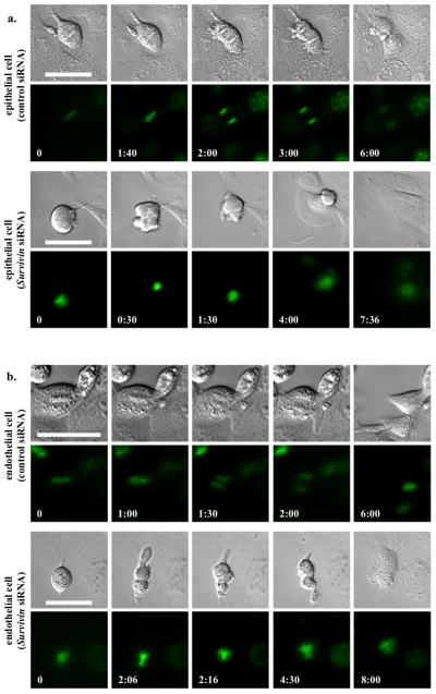
Methods and results: Cysts and aneurysms from polycystic kidney disease patients, Pkd mouse, and zebrafish models are characterized by chromosome instability and low survivin expression. This triggers cytokinesis defects and formation of nuclear polyploidy or aneuploidy. In vivo conditional mouse and zebrafish models confirm that survivin gene deletion in the kidneys results in a cystic phenotype. As in hypertensive Pkd1, Pkd2, and Tg737 models, aneurysm formation can also be induced in vascular-specific normotensive survivin mice. Survivin knockout also contributes to abnormal oriented cell division in both kidney and vasculature. Furthermore, survivin expression and ciliary localization are regulated by flow-induced cilia activation through protein kinase C, Akt and nuclear factor-κB. Circumventing ciliary function by re-expressing survivin can rescue polycystic kidney disease phenotypes.

Conclusions: For the first time, our studies offer a unifying mechanism that explains both renal and vascular phenotypes in polycystic kidney disease. Although primary cilia dysfunction accounts for aneurysm formation and hypertension, hypertension itself does not cause aneurysm. Furthermore, aneurysm formation and cyst formation share a common cellular and molecular pathway involving cilia function or structure, survivin expression, cytokinesis, cell ploidy, symmetrical cell division, and tissue architecture orientation.

Keywords: aurora kinase; blood flow; blood pressure; cardiovascular system; endothelium, vascular; epithelium.


Conflict of interest statement

Conflict of Interest Disclosures: None.

Figures

Figure 1
Figure 1
Autosomal dominant polycystic kidney disease (ADPKD) renal epithelia are characterized by abnormal ploidy level and survivin down-regulation. (a) Karyotyping was carried out in freshly isolated epithelial cells from non-ADPKD patients to visualize individual chromosomes (non-cytic kidney). (b) Characterization of individual chromosomes from a single renal epithelium isolated from ADPKD patient indicated tetraploid and abnormal chromosomal composition. (c) Overall karyotype analysis of individual cells confirmed the presence of abnormal genomic compositions (aneuploidy or polyploidy) in cells from ADPKD patients. (d) Kidney tissues from ADPKD patients were also used to confirm survivin expression, and GADPH was used as loading control. Bar graph shows relative survivin expression levels. N=3 each for freshly isolated non-ADPKD and ADPKD kidneys.
Figure 2
Figure 2
Survivin downregulation is sufficient to induce cystic kidney formation. (a-i) Unilateral ureteral obstruction (UUO) was performed on either wild-type or Survivin knockout (Mx1Cre:Survivinflox/flox) mice at one-month old, and mice were sacrificed a week later. (a-ii) Mx1Cre:Survivinflox/flox induced- and non-induced littermates were sacrificed, and their kidneys were compared three months later. Comparison of gross features revealed enlargement in Survivin knockout kidneys with an apparent bulged and fluid-filled kidney. (b) H&E-stained kidney sections from five-week old wild-type as well as Mx1Cre:survivinflox/flox mice with or without UUO surgery are shown. Florescence studies were performed with DBA staining (red) and counterstained with WGA (green) and DAPI (blue).
Figure 3
Figure 3
Aneurysm formation is an extra-renal phenotype of PKD. (a) Aneurysm surgery was performed in mice at one-month-old and mice were sacrificed at three months of age. The aortas were isolated and their diameters were measured at the surgical site. Unlike wild-type, Pdgfβcre:Survivinflox/flox mice showed a severe aneurysm induction to a similar extent shown in Pdgfβcre:Pkd1flox/flox, Pkd2+/−, and Tg737orpk/orpk mice. (b) Representatives of cross-sections of aortas at the aneurysm surgery site are shown in control wild-type, Pdgfβcre:Pkd1flox/flox, Pkd2+/−, Tg737orpk/orpk, or Pdgfβcre:Survivinflox/flox mice with and without aneurysm surgery. Similar to Pdgfβcre:Pkd1flox/flox, Pkd2+/− and Tg737orpk/orpk mice, Pdgfβcre:Survflox/flox mice exhibited aneurysm formation and aortic dilation compared to wild-type mice. Arrows point to aneurysm formation, and double head arrows point to aorta diameter. N≥3 for each group and genotype. Bar=200 μm. (c) Bar graph shows averaged values for aorta diameter in wild-type, Pdgfβcre:Pkd1flox/flox, Pkd2+/−, Tg737orpk/orpk, or Pdgfβcre:Survivinflox/flox mice. (d) The grade of aneurysm is also tabulated.
Figure 4
Figure 4
Survivin down-regulation is associated with abnormal cytokinesis in primary cells of renal epithelia and vascular endothelial cells. (a) To obtain the mechanistic insights of cytokinesis defect in renal epithelial cells, live-cell imaging analysis was performed. (b) A similar study was also done in vascular endothelial cells. Control and survivin knockdown cells were loaded with Hoechst dye to examine nuclear division (lower panels), while DIC images were used to study cytokinesis (top panels). Time stamps indicate hours and minutes as illustrated in Supplementary Materials, Movies S1–S4. Bar=50 μm. N≥3 for each group and treatment.
Figure 5
Figure 5
Abnormal cellular division orientation is associated with renal cystic and vascular aneurysm phenotypes. (a) Kidney tubular sections from both Mx1Cre:survivinflox/flox mice, with or without UUO surgery, showed abnormal cell division and division orientation with respect to the axis of the kidney tubule. ZO-1 staining was used to indicate renal tubule orientation, and cell-undergoing division within the region is further enlarged. (b) Longitudinal abdominal aortic sections in non-surgery (control) and aneurysm-induced (surgery) models were studied to analyze endothelial orientation and cell division. Nucleus from smooth muscle cells was shown in blue, and nucleus of a single intimal layer of endothelial tissue was pseudo-colored in yellow. Abnormal randomized cell orientation is clearly visible. In all figures, division orientation relative to tubule/artery axis is shown in green double head arrows, and abnormal cell division is indicated in green arrows. N≥3 for each group and genotype. N≥100 for distribution of spindle orientation angle for each genotype and each treatment. Bar=40 μm.
Figure 6
Figure 6
Cilia regulate cell division through survivin expression. (a) Western blot analysis was used to study survivin and aurora-A expressions in wild-type and cilia mutant cells (Pkd1−/− and Tg737Orpk/Orpk) in the presence or absence of fluid-shear stress. GAPDH and actin were used as loading controls. Bar graph represents averaged survivin and aurora-A expressions. (b) Acetylated-α-tubulin was used as a ciliary marker to indicate ciliary expression and localization of survivin in response to fluid-shear in wild-type but not mutant cells. (c) Cells treated with survivin or aurora-A inhibitors are characterized by multiple centrosomes, abnormal mitotic spindle, and mitotic arrest during cell division. Cells were stained with acetylated-α-tubulin (green) and pericentrin (red) and captured at resting (i) and dividing (ii) stages of cell cycle. Bar=10 μm. N=3 for each cell type and treatment. Statistics was performed by comparing individual group to their corresponding wild-type static control groups.
Figure 7
Figure 7
PKC/Akt/NF-κB signaling pathway regulates flow-induced survivin expression and cell division. (a) After wild-type and cilia mutant (Pkd1−/− and Tg737Orpk/Orpk) cells were treated with PKC inhibitor or activator, both Akt and p-Akt were analyzed. When treated with PKC inhibitor, all cell lines showed down-regulation of p-Akt, while PKC activator treatment showed an increase in p-Akt compared to non-treated control cells. (b) The effect of fluid-flow on Akt and aurora-A expression was analyzed in the presence or absence of PKC inhibitor. When subjected to fluid-shear, p-Akt expression was up-regulated only in wild-type cells. While p-Akt expression returned to basal levels following treatment with PKC inhibitor and fluid-shear stress in wild-type cells, it stayed repressed in mutant cells. (c) Treatment with aurora-A inhibitor resulted in a decrease in p-Akt, aurora-A and survivin expression; however, these decreases were not significant from the control, non-treated group. (d) While total Akt level was not changed, fluid-shear stress significantly induced expression of p-Akt in wild-type but not in mutant cells. Aurora-A expression was increased following fluid-shear stress in wild-type cells; however, this increase was not significant from control. Both NF-κB and pNF-κB expressions were increased following fluid-shear stress only in wild-type cells, while mutant cells maintained a high basal level of NF-κB compared to static wild-type cells. Survivin expression was increased following shear-stress in wild-type cells. (e) Western blot analyses were conducted to confirm the signaling mechanism involving survivin expression by siRNA-mediated knockdown of PKC, Akt, aurora A, or survivin. To further confirm the involvement of these signaling molecules in centrosome number and cell division abnormality, immunofluorescence and flow cytometry analyses were presented in the Supplementary Materials together with the statistics.
Figure 8
Figure 8
Survivin overexpression rescued PKD phenotypes in zebrafish. (a) Zebrafish embryos were scored for phenotypic observations at different developmental stages. Shown here are representative images of zebrafish at 24, 48 and 72 hours-post-fertilization (hpf) injected with either control morpholino (MO), pkd2 MO, pkd2 MO plus survivin mRNA, survivin mRNA alone, or pkd2 MO plus VEGF. Abnormal phenotypes associated with pkd2 MO injections such as curly tail and renal cyst were rescued by survivin mRNA or VEGF injection into zebrafish embryos. Representative images of 48-hpf zebrafish sections are shown. The sections were stained with H&E (bottom panels), and arrows point to pronephric structures. (b) RT-PCR was performed to examine zebrafish (zf) and human (hu) transcript levels for survivin and to confirm pkd2 knockdown. Human survivin was introduced through mRNA injection. α-tubulin was used as a loading control. (c) Expression levels of survivin were analyzed in all the groups, in which zebrafish and human survivin can be recognized by the same antibody. (d) Analyses of individual chromosomes were performed in all groups of fish embryos to study chromosomal number. Black bar=500 μm; red bar=50 μm. All quantification and statistical analyses on Western blot, RT-PCR and polyploidy level are presented in the Supplementary Materials.

References

    1. AbouAlaiwi WA, Takahashi M, Mell BR, Jones TJ, Ratnam S, Kolb RJ, Nauli SM. Ciliary polycystin-2 is a mechanosensitive calcium channel involved in nitric oxide signaling cascades. Circ Res. 2009;104:860–9. - PMC - PubMed
    1. Nauli SM, Alenghat FJ, Luo Y, Williams E, Vassilev P, Li X, Elia AE, Lu W, Brown EM, Quinn SJ, Ingber DE, Zhou J. Polycystins 1 and 2 mediate mechanosensation in the primary cilium of kidney cells. Nat Genet. 2003;33:129–37. - PubMed
    1. Nauli SM, Kawanabe Y, Kaminski JJ, Pearce WJ, Ingber DE, Zhou J. Endothelial cilia are fluid shear sensors that regulate calcium signaling and nitric oxide production through polycystin-1. Circulation. 2008;117:1161–71. - PMC - PubMed
    1. Xu C, Shmukler BE, Nishimura K, Kaczmarek E, Rossetti S, Harris PC, Wandinger-Ness A, Bacallao RL, Alper SL. Attenuated, flow-induced ATP release contributes to absence of flow-sensitive, purinergic Cai2+ signaling in human ADPKD cyst epithelial cells. Am J Physiol Renal Physiol. 2009;296:F1464–76. - PMC - PubMed
    1. AbouAlaiwi WA, Ratnam S, Booth RL, Shah JV, Nauli SM. Endothelial cells from humans and mice with polycystic kidney disease are characterized by polyploidy and chromosome segregation defects through survivin down-regulation. Hum Mol Genet. 2011;20:354–67. - PMC - PubMed

Publication types

MeSH terms