

. 2013 Sep 21;5(18):10.1039/C3AY41124A.
doi: 10.1039/C3AY41124A.
Electron Ionization-Induced Release of Coded Isotopic Reporter Ions in an m/z Zone of Minimal Interference for Quantifiable, Multiplexed GC-MS Analyses
Affiliations
- PMID: 24235976
- PMCID: PMC3822575
- DOI: 10.1039/C3AY41124A
Item in Clipboard
Electron Ionization-Induced Release of Coded Isotopic Reporter Ions in an m/z Zone of Minimal Interference for Quantifiable, Multiplexed GC-MS Analyses
Anal Methods.
.
Abstract
We describe an isotope coding strategy that enables simultaneous GC-MS analysis of multiple samples for substrate identification and quantification. The method relies on direct measurement of isotopic ethyl carbenium ions serving as mass spectral tags in a zone of minimal interference (ZMI) at m/z 31-37. Sample aldehyde and ketone mixtures were reacted with isotopic 2-aminooxyethyl propionates to illustrate the method, which determined the relative abundance of the mixed compounds with an average 95% accuracy. ZMI reporter ion detection also enables chemoselective substrate profiling and absolute quantification, as demonstrated using a biologically derived sample.
Figures

Representation of a multiplexed GC-MS analysis using isotope-coded reagents designed for electron-induced expulsion of reporter mass spectral tags (MSTs). The derivatization reagents provide corresponding MSTs for relative quantification of substrates from separate sample mixtures.

Delineation of the zone of minimal interference; A: fragment ion frequency for the compound library (NIST/EPA/NIH NIST 08 Mass Spectral Library), B: corresponding summed ion count (peak intensity) for a given m/z.

ZMI plots obtained from the eluted peaks of the pooled sample mixture of Scheme 3 showing the MST ion abundance (vertical) for the range m/z 32–34. Shown are the mean values of three separate experiments and the standard deviation from the mean. The starting A:B:C substrate ratio is given in brackets. Ion counts are normalized to 100 for the parent fragments (not shown).

A. GCxGC-TOF-MS total ion chromatogram (TIC) of turmeric root extract after derivatization using AEP reagents; B. Extracted ion chromatogram (EIC) for peaks that exhibit MS signals at m/z 33 and 34 (same field of view as in A with lower threshold); C. Expanded field of view of peaks within the highlighted rectangle of B where circled peaks 1–6 are, respectively, the AEP-adducts of 2-nonanone, 2-decanone, 2-undecanone, 2-dodecanone, 2-tridecanone and 2-octadecanone.

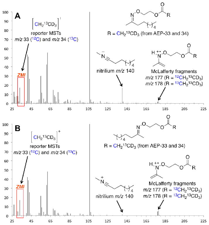
Characteristic EI-MS fragmentations of ketoxime ethers.
EI-Mass spectra of AEP–2-nonanone adducts: (A) Z-isomer, (B) E-isomer. Adducts were obtained by derivatization of turmeric root extract using AEP-33 and AEP-34 in a 1:1 ratio.

EI-MS–induced fragmentation of AEP derived oxime ether adducts.

Synthesis of AEP reagents. Reagents: (a) CD3CH2CO2H (to 5a) or 13CD3CH2CO2H (5b) or 13CD313CH2CO2H (5c), DIC (1.5 eq), DMAP (cat.), CH2Cl2, rt; (b) MeNHNH2 (1.1 eq), CH2Cl2, 0 °C, 45 min.

Formulation, labeling and pooling of carbonyl sample mixtures for analysis. Depicted at bottom is the total ion chromatogram of the pooled sample mixture showing elution of the oxime ether adducts of the substrates indicated by name.
References
-
- Almstetter MF, Oefner PJ, Dettmer K. Anal Bioanal Chem. 2012;402:1993–2013. - PubMed
-
- Wei R, Li G, Seymour AB. Anal Chem. 2010;82:5527–5533. - PubMed
-
- Courant F, Pinel G, Bichon E, Monteau F, Antignac JP, Le Bizec B. Analyst. 2009;134:1637–1646. - PubMed
- Lv H, Palacios G, Hartil K, Kurland IJ. J Proteome Res. 2011;10:2104–2112. - PMC - PubMed
- Aoki M, Konya Y, Takagaki T, Umemura K, Sogame Y, Katsumata T, Komuro S. Rapid Commun Mass Spectrom. 2011;25:1847–1852. - PubMed
- Spagou K, Wilson ID, Masson P, Theodoridis G, Raikos N, Coen M, Holmes E, Lindon JC, Plumb RS, Nicholson JK, Want EJ. Anal Chem. 2011;83:382–390. - PubMed
- Xiao JF, Zhou B, Ressom HW. Trends Anal Chem. 2012;32:1–14. - PMC - PubMed
Grants and funding
LinkOut - more resources
Full Text Sources
Other Literature Sources
Miscellaneous