. 2013 Nov 13;8(11):e79350.
doi: 10.1371/journal.pone.0079350. eCollection 2013.

Lipopolysaccharide induces degradation of connexin43 in rat astrocytes via the ubiquitin-proteasome proteolytic pathway

Affiliations

Lipopolysaccharide induces degradation of connexin43 in rat astrocytes via the ubiquitin-proteasome proteolytic pathway

Chih-Kai Liao et al. PLoS One. .

Abstract

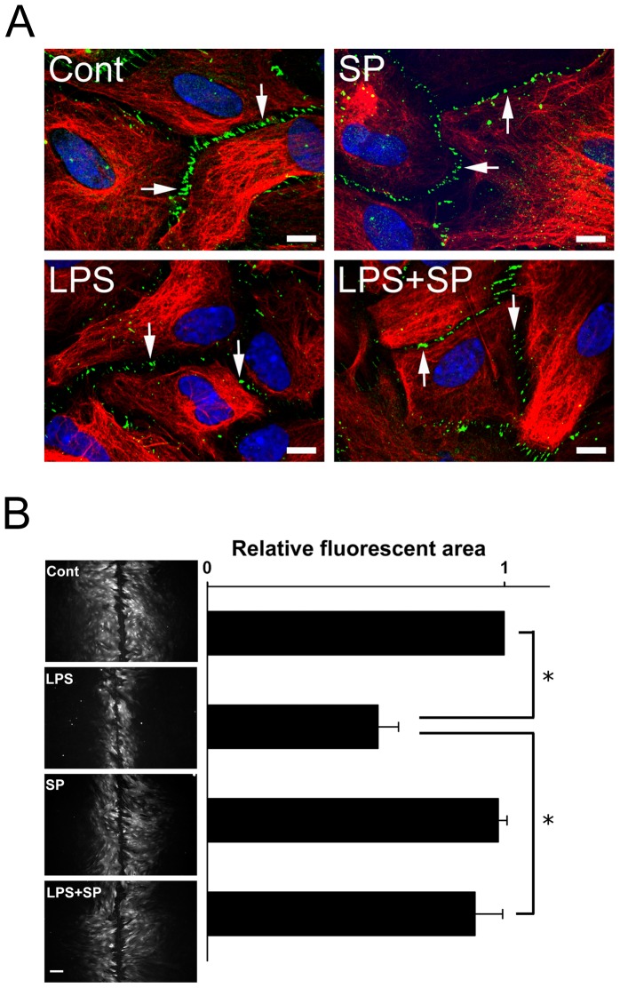
The astrocytic syncytium plays a critical role in maintaining the homeostasis of the brain through the regulation of gap junction intercellular communication (GJIC). Changes to GJIC in response to inflammatory stimuli in astrocytes may have serious effects on the brain. We have previously shown that lipopolysaccharide (LPS) reduces connexin43 (Cx43) expression and GJIC in cultured rat astrocytes via a toll-like receptor 4-mediated signaling pathway. In the present study, treatment of astrocytes with LPS resulted in a significant increase in levels of the phosphorylated forms of stress-activated protein kinase/c-Jun N-terminal kinase (SAPK/JNK) -1, -2, and -3 for up to 18 h. An increase in nuclear transcription factor NF-κB levels was also observed after 8 h of LPS treatment and was sustained for up to 18 h. The LPS-induced decrease in Cx43 protein levels and inhibition of GJIC were blocked by the SAPK/JNK inhibitor SP600125, but not by the NF-κB inhibitor BAY11-7082. Following blockade of de novo protein synthesis by cycloheximide, LPS accelerated Cx43 degradation. Moreover, the LPS-induced downregulation of Cx43 was blocked following inhibition of 26S proteasome activity using the reversible proteasome inhibitor MG132 or the irreversible proteasome inhibitor lactacystin. Immunoprecipitation analyses revealed an increased association of Cx43 with both ubiquitin and E3 ubiquitin ligase Nedd4 in astrocytes after LPS stimulation for 6 h and this effect was prevented by SP600125. Taken together, these results suggest that LPS stimulation leads to downregulation of Cx43 expression and GJIC in rat astrocytes by activation of SAPK/JNK and the ubiquitin-proteasome proteolytic pathway.


Conflict of interest statement

Competing Interests: The authors have declared that no competing interests exist.

Figures

Figure 1
Figure 1. Time-dependent effects of LPS on levels of NF-κB, Cx43, and phospho-JNK in rat astrocytes.
(A) Astrocytes were treated with 2 µg/ml of LPS for 0, 6, 8, 10, 12, 14, or 18 h, then whole cell lysates were collected, subjected to10% SDS-PAGE, and analyzed by immunoblotting with antibodies against NF-κB (NF-κB) or total Cx43 (Cx43). Glyceraldehyde 3-phosphate dehydrogenase (GAPDH) was used as the loading control. The three major bands of Cx43 are the non-phosphorylated form (P0) and the two phosphorylated forms (P1 and P2). (B) The blots from 4 independent experiments were subjected to densitometric analyses for total Cx43 (P0 plus P1 plus P2) and the results expressed as the density of the bands in the test sample relative to those in the time zero sample. *P<0.05, **P<0.01 compared to 0 h using Dunnett’s post-hoc test. (C) Whole cell lysates were prepared from astrocytes treated with 2 µg/ml LPS for 0, 1, 3, 6, or 18 h, subjected to 10% SDS-PAGE and immunoblotted with antibodies against the three isoforms (JNK1, JNK2, and JNK3) of phospho-JNK(pJNK) or GAPDH (GAPDH, loading control). (D) The blots from 3 independent experiments were subjected to densitometric analyses for phospho-pJNK (pJNK1 plus pJNK2/3) and the results expressed as the density of the bands in the test sample relative to those in the time zero sample. *P<0.05, **P<0.01 compared to 0 h using Dunnett’s post-hoc test.
Figure 2
Figure 2. The JNK inhibitor SP600125 inhibits LPS-induced downregulation of Cx43 in astrocytes.
(A) Astrocytes were treated for 18 h with 0.05% DMSO (Cont), 2 µg/ml of LPS (LPS), 10 µM SP600125 (SP), or 5 µM BAY11-7082 (BAY) or were treated with SP600125 (LPS+SP) or BAY11-7082 (LPS+BAY) for 30 min prior to, and during, incubation with LPS for 18 h. Cell lysates were prepared and total proteins analyzed by 10% SDS-PAGE and immunoblotting using antibodies against total Cx43 or GAPDH (GAPDH, loading control). (B) The blots from 3 independent experiments were subjected to densitometric analyses for total Cx43 (P0 plus P1 plus P2) and the results expressed as the density of the bands in the test sample relative to those in the control. *P<0.01, NS (not significant) compared to the LPS-treated group using Dunnett’s post-hoc test.
Figure 3
Figure 3. Effects of SP600125 on Cx43 gap junction plaque staining and 6-CF fluorescent dye transfer in LPS-treated rat astrocytes.
(A) Astrocytes were treated for 18 h with 0.05% DMSO (Cont), 2 µg/ml of LPS (LPS), 10 µM SP600125 (SP), or 5 µM BAY11-7082 (BAY) or were treated with SP600125 (LPS+SP) or BAY11-7082 (LPS+BAY) for 30 min prior to, and during, incubation with LPS for 18 h. The confocal microscopy merged images show control or treated astrocytes double-immunostained with antibodies against Cx43 (Cx43, green) and glial fibrillary acidic protein (GFAP, red). Nuclei were stained with DAPI (blue). The arrows indicate Cx43 gap junction plaques at cell-cell contact points. Bar = 10 µm. (B) Astrocytes were treated for 18 h with 0.05% DMSO (Cont) or 2 µg/ml of LPS (LPS) or were preteated for 30 min with 10 µM SP600125 followed by treatment for 18 h in absence of LPS (SP) or were pretreated for 30 min with SP600125 followed by LPS treatment for 18 h in the continued presence of the inhibitor (LPS+SP) subjected to 6-CF fluorescent dye scrape loading as described in the Materials and Methods (left panels, bar = 100 µm). The right panel shows the data for the dye-spreading area from 3 independent experiments expressed as the fluorescent area relative to that in the control. *P<0.01 compared to the LPS-treated group using Dunnett’s post-hoc test.
Figure 4
Figure 4. LPS accelerates Cx43 degradation in cycloheximide-treated astrocytes.
(A) Astrocytes were pretreated with 2 µg/ml LPS (LPS) for 8 h followed by treatment for 1, 3, or 5 h with 10 µg/ml of cycloheximide in the absence (CHX) or in the continued presence of LPS (LPS+CHX), then the cells were lysed and the whole cell lysates subjected to10% SDS-PAGE, and analyzed by immunoblotting with antibodies against total Cx43 (Cx43, P0, P1, and P2). The blots were then stripped and reprobed with antibodies against GAPDH (GAPDH, loading control). (B) The blots from 3 independent experiments were subjected to densitometric analyses for total Cx43 (P0 plus P1 plus P2) and the results expressed as the density of the bands in the test sample relative to those in the time zero sample of CHX or LPS+CHX. *P<0.05, **P<0.01 compared to CHX using independent-sample t-test.
Figure 5
Figure 5. SP600125 prevents Cx43 ubiquitination in LPS-treated rat astrocytes.
(A) Astrocytes were treated with 2 µg/ml of LPS for 0, 3, 6, 8, or 10 h. The cell lysates immunopreciptated using normal rabbit serum (NRS, immunoprecipitation control, IPC) or rabbit antibody against total Cx43 (IP: Cx43). The immunoprecipitates were then subjected to 10% SDS-PAGE and immunoblotting using antibodies against ubiquitin (IB: Ubi). The immunoprecipitates were then subjected to 10% SDS-PAGE and immunoblotting using antibodies against ubiquitin (IB: Ubi) or total Cx43 (IB: Cx43). (B) The blots from 3 independent experiments were subjected to densitometric analyses for ubiquitinated Cx43 and the results expressed as the density of the bands in the test sample relative to those in the time zero sample. *P<0.05, NS (not significant) compared to 0 h using Dunnett’s post-hoc test. (C) The cell lysates from astrocytes treated with 0.05% DMSO (Cont), 2 µg/ml of LPS (LPS) for 6 h, or were pretreated for 30 min with 10 µM SP600125 followed by LPS (LPS+SP) treatment for 6 h in the presence of the inhibitor were immunopreciptated using normal rabbit serum (NRS, immunoprecipitation control, IPC) or rabbit antibody against total Cx43 (IP: Cx43). The immunoprecipitates were then subjected to 10% SDS-PAGE and immunoblotting using antibodies against ubiquitin (IB: Ubi), Nedd4 (IB: Nedd4) or total Cx43 (IB: Cx43). (D) The blots from 3 independent experiments were subjected to densitometric analyses for Nedd4 or ubiquitinated Cx43 and the results expressed as the density of the bands in the test sample relative to those in the control. *P<0.05, **P<0.01 compared to the LPS-treated group using Dunnett’s post-hoc test.
Figure 6
Figure 6. Effect of the proteasome inhibitor MG132 on Cx43 levels in LPS-treated astrocytes.
(A) Astrocytes were treated with 0.05% DMSO for 8 h (Cont), with 5 µM MG132 for 4, 6, or 8 h (M4, M6, M8), with 2 µg/ml of LPS for 18 h (LPS), or with LPS for 18 h with addition of MG132 for the last 4, 6, or 8 h (LPS+M4, LPS+M6, LPS+M8), then cell lysates were prepared, subjected to 10% SDS-PAGE, and analyzed by immunoblotting with antibodies against total Cx43 (Cx43) or GAPDH (GAPDH, loading control). (B) Densitometric analyses of total Cx43 (P0 plus P1 plus P2) from 3 independent experiments expressed as the density of the bands in the test sample relative to those in the control. *P<0.01, NS (not significant) compared to the LPS-treated group using Dunnett’s post-hoc test.
Figure 7
Figure 7. Effect of the proteasome inhibitor lactacystin on Cx43 levels in LPS-treated astrocytes.
(A) Astrocytes were treated for 8 h with 0.1% DMSO (Cont), for 18 h with 2 µg/ml of LPS (LPS), for 8 h with 5 µM lactacystin (Lact), or with 2 µg/ml LPS for 18 h with addition of lactacystin for the last 8 h (LPS+Lact), then cell lysates were prepared, subjected to 10% SDS-PAGE, and analyzed by immunoblotting with antibodies against total Cx43 (Cx43, P0, P1, and P2) or GAPDH (GAPDH, loading control). (B) Densitometric analyses of total Cx43 (P0 plus P1 plus P2) from 3 independent experiments expressed as the density of the bands in the test sample relative to those in the control. **P<0.01, *P<0.05 compared to the LPS-treated groups using Dunnett’s post-hoc test.
Figure 8
Figure 8. Schematic illustration of the signaling pathway involved in the inhibitory effect of LPS on Cx43 gap junctions in rat astrocytes.
LPS treatment down-regulates caveolin-3 levels via TLR4-mediated iNOS activation, which in turn reduces the expression of Cx43. LPS also induces JNK phosphorylation, resulting in GJIC inhibition, gap junction disassembly, and Cx43 degradation by the ubiquitin-proteasome proteolytic pathway.

Similar articles

Cited by

References

    1. Giaume C, Koulakoff A, Roux L, Holcman D, Rouach N (2010) Astroglial networks: a step further in neuroglial and gliovascular interactions. Nat Rev Neurosci 11: 87–99. - PubMed
    1. Giaume C, Theis M (2010) Pharmacological and genetic approaches to study connexin-mediated channels in glial cells of the central nervous system. Brain Res Rev 63: 160–176. - PubMed
    1. Dermietzel R, Hertberg EL, Kessler JA, Spray DC (1991) Gap junctions between cultured astrocytes: immunocytochemical, molecular, and electrophysiological analysis. J Neurosci 11: 1421–1432. - PMC - PubMed
    1. Dermietzel R, Gao Y, Scemes E, Vieira D, Urban M, et al. (2000) Connexin43 null mice reveal that astrocytes express multiple connexins. Brain Res Brain Res Rev 32: 45–56. - PubMed
    1. Kielian T (2008) Glial connexins and gap junctions in CNS inflammation and disease. J Neurochem 106: 1000–1016. - PMC - PubMed

Publication types

MeSH terms