

Male killing Spiroplasma preferentially disrupts neural development in the Drosophila melanogaster embryo
- PMID: 24236124
- PMCID: PMC3827344
- DOI: 10.1371/journal.pone.0079368
Male killing Spiroplasma preferentially disrupts neural development in the Drosophila melanogaster embryo
Abstract
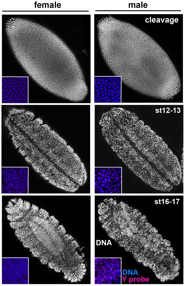
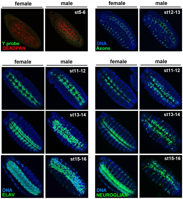
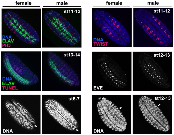
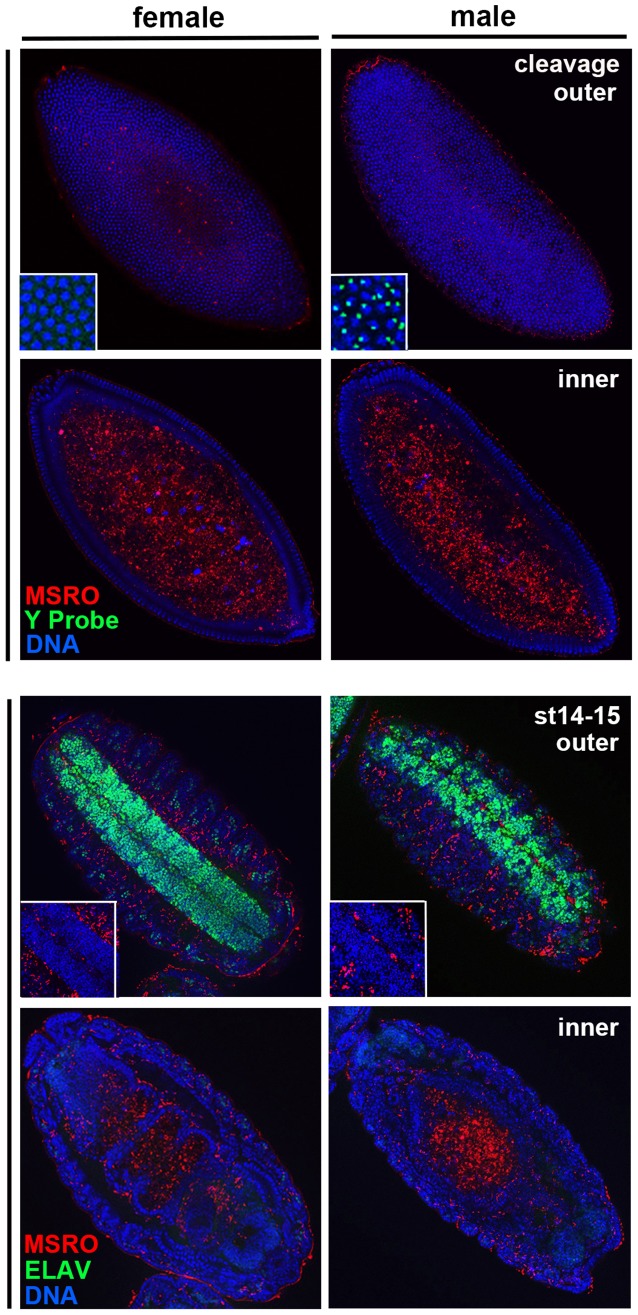
Male killing bacteria such as Spiroplasma are widespread pathogens of numerous arthropods including Drosophila melanogaster. These maternally transmitted bacteria can bias host sex ratios toward the female sex in order to 'selfishly' enhance bacterial transmission. However, little is known about the specific means by which these pathogens disrupt host development in order to kill males. Here we show that a male-killing Spiroplasma strain severely disrupts nervous tissue development in male but not female D. melanogaster embryos. The neuroblasts, or neuron progenitors, form properly and their daughter cells differentiate into neurons of the ventral nerve chord. However, the neurons fail to pack together properly and they produce highly abnormal axons. In contrast, non-neural tissue, such as mesoderm, and body segmentation appear normal during this time, although the entire male embryo becomes highly abnormal during later stages. Finally, we found that Spiroplasma is altogether absent from the neural tissue but localizes within the gut and the epithelium immediately surrounding the neural tissue, suggesting that the bacterium secretes a toxin that affects neural tissue development across tissue boundaries. Together these findings demonstrate the unique ability of this insect pathogen to preferentially affect development of a specific embryonic tissue to induce male killing.
Conflict of interest statement
Figures
Similar articles
-
Effect of heritable symbionts on maternally-derived embryo transcripts.Sci Rep. 2019 Jun 20;9(1):8847. doi: 10.1038/s41598-019-45371-0. Sci Rep. 2019. PMID: 31222094 Free PMC article.
-
Male-Killing Spiroplasma Alters Behavior of the Dosage Compensation Complex during Drosophila melanogaster Embryogenesis.Curr Biol. 2016 May 23;26(10):1339-45. doi: 10.1016/j.cub.2016.03.050. Epub 2016 May 5. Curr Biol. 2016. PMID: 27161498 Free PMC article.
-
Genome sequence of the Drosophila melanogaster male-killing Spiroplasma strain MSRO endosymbiont.mBio. 2015 Mar 31;6(2):e02437-14. doi: 10.1128/mBio.02437-14. mBio. 2015. PMID: 25827421 Free PMC article.
-
The Spiroplasma heritable bacterial endosymbiont of Drosophila.Fly (Austin). 2010 Jan-Mar;4(1):80-7. doi: 10.4161/fly.4.1.10883. Epub 2010 Jan 5. Fly (Austin). 2010. PMID: 20081357 Review.
-
[Symbiotic bacteria, which modify reproduction processes of Drosophila melanogaster].Mikrobiol Z. 2011 Mar-Apr;73(2):43-52. Mikrobiol Z. 2011. PMID: 21598659 Review. Russian.
Cited by
-
Effect of heritable symbionts on maternally-derived embryo transcripts.Sci Rep. 2019 Jun 20;9(1):8847. doi: 10.1038/s41598-019-45371-0. Sci Rep. 2019. PMID: 31222094 Free PMC article.
-
Genome elimination mediated by gene expression from a selfish chromosome.Sci Adv. 2020 Apr 3;6(14):eaaz9808. doi: 10.1126/sciadv.aaz9808. eCollection 2020 Apr. Sci Adv. 2020. PMID: 32284986 Free PMC article.
-
Male-killing toxin in a bacterial symbiont of Drosophila.Nature. 2018 May;557(7704):252-255. doi: 10.1038/s41586-018-0086-2. Epub 2018 May 2. Nature. 2018. PMID: 29720654 Free PMC article.
-
Male-killing symbiont damages host's dosage-compensated sex chromosome to induce embryonic apoptosis.Nat Commun. 2016 Sep 21;7:12781. doi: 10.1038/ncomms12781. Nat Commun. 2016. PMID: 27650264 Free PMC article.
-
Sex determination systems as the interface between male-killing bacteria and their hosts.Proc Biol Sci. 2022 Apr 13;289(1972):20212781. doi: 10.1098/rspb.2021.2781. Epub 2022 Apr 13. Proc Biol Sci. 2022. PMID: 35414231 Free PMC article. Review.
References
-
- Whitcomb RF (1980) The Genus Spiroplasma. Annual Review of Microbiology 34: 677–709. - PubMed
-
- Haselkorn TS (2010) The Spiroplasma heritable bacterial endosymbiont of Drosophila. Fly 4: 80–87. - PubMed
-
- Hurst GDD, Jiggins FM, von der Schulenburg JHG, Bertrand D, West SA, et al. (1999) Male-killing Wolbachia in two species of insect. Proceedings of the Royal Society B-Biological Sciences 266: 735–740.
MeSH terms
LinkOut - more resources
Full Text Sources
Other Literature Sources
Molecular Biology Databases
