

AUTOMATED ANATOMICAL LABELING OF THE CEREBRAL ARTERIES USING BELIEF PROPAGATION
- PMID: 24236229
- PMCID: PMC3824264
- DOI: 10.1117/12.2006460
AUTOMATED ANATOMICAL LABELING OF THE CEREBRAL ARTERIES USING BELIEF PROPAGATION
Abstract
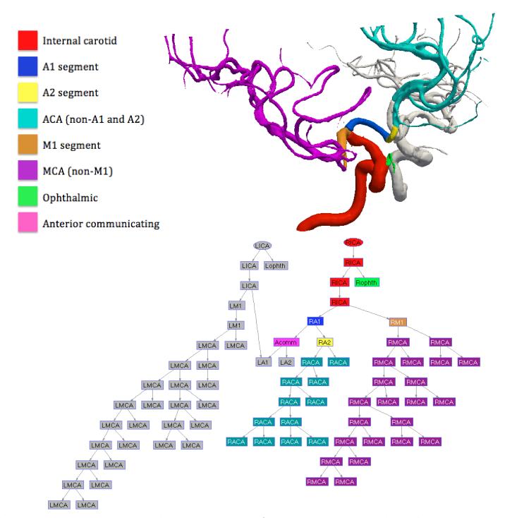
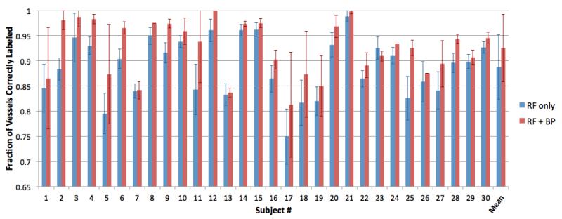
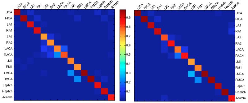
Labeling of cerebral vasculature is important for characterization of anatomical variation, quantification of brain morphology with respect to specific vessels, and inter-subject comparisons of vessel properties and abnormalities. We propose an automated method to label the anterior portion of cerebral arteries using a statistical inference method on the Bayesian network representation of the vessel tree. Our approach combines the likelihoods obtained from a random forest classifier trained using vessel centerline features with a belief propagation method integrating the connection probabilities of the cerebral artery network. We evaluate our method on 30 subjects using a leave-one-out validation, and show that it achieves an average correct vessel labeling rate of over 92%.
Keywords: Automated labeling of vessels; belief propagation; cerebral arteries; random forest; statistical inference on Bayesian networks.
Figures
Similar articles
-
Automated intracranial vessel labeling with learning boosted by vessel connectivity, radii and spatial context.Proc Mach Learn Res. 2022 Nov;194:34-44. Proc Mach Learn Res. 2022. PMID: 37077315 Free PMC article.
-
Anatomical labeling of the anterior circulation of the Circle of Willis using maximum a posteriori classification.Med Image Comput Comput Assist Interv. 2011;14(Pt 3):330-7. doi: 10.1007/978-3-642-23626-6_41. Med Image Comput Comput Assist Interv. 2011. PMID: 22003716
-
Automated Method for Retinal Artery/Vein Separation via Graph Search Metaheuristic Approach.IEEE Trans Image Process. 2019 Jan 1. doi: 10.1109/TIP.2018.2889534. Online ahead of print. IEEE Trans Image Process. 2019. PMID: 30605099
-
Automatic anatomical labeling of arteries and veins using conditional random fields.Int J Comput Assist Radiol Surg. 2017 Jun;12(6):1041-1048. doi: 10.1007/s11548-017-1549-x. Epub 2017 Mar 8. Int J Comput Assist Radiol Surg. 2017. PMID: 28275889
-
Common features of the cerebral perforating arteries and their clinical significance.Acta Neurochir (Wien). 2015 May;157(5):743-54; discussion 754. doi: 10.1007/s00701-015-2378-8. Epub 2015 Mar 14. Acta Neurochir (Wien). 2015. PMID: 25772345 Review.
Cited by
-
An Improved Path-Finding Method for the Tracking of Centerlines of Tortuous Internal Carotid Arteries in MR Angiography.J Imaging. 2024 Feb 28;10(3):58. doi: 10.3390/jimaging10030058. J Imaging. 2024. PMID: 38535138 Free PMC article.
-
Automated anatomical labeling of coronary arteries via bidirectional tree LSTMs.Int J Comput Assist Radiol Surg. 2019 Feb;14(2):271-280. doi: 10.1007/s11548-018-1884-6. Epub 2018 Nov 27. Int J Comput Assist Radiol Surg. 2019. PMID: 30484116
-
MR Imaging of Human Brain Mechanics In Vivo: New Measurements to Facilitate the Development of Computational Models of Brain Injury.Ann Biomed Eng. 2021 Oct;49(10):2677-2692. doi: 10.1007/s10439-021-02820-0. Epub 2021 Jul 1. Ann Biomed Eng. 2021. PMID: 34212235 Free PMC article. Review.
-
TTN: Topological Transformer Network for Automated Coronary Artery Branch Labeling in Cardiac CT Angiography.IEEE J Transl Eng Health Med. 2023 Nov 1;12:129-139. doi: 10.1109/JTEHM.2023.3329031. eCollection 2024. IEEE J Transl Eng Health Med. 2023. PMID: 38074924 Free PMC article.
-
Automated in-depth cerebral arterial labelling using cerebrovascular vasculature reframing and deep neural networks.Sci Rep. 2023 Feb 24;13(1):3255. doi: 10.1038/s41598-023-30234-6. Sci Rep. 2023. PMID: 36828857 Free PMC article.
References
-
- Pérez-Carrillo GJG, Hogg JP. Intracranial vascular lesions and anatomical variants all residents should know. Current Problems in Diagnostic Radiology. 2010;39(3):91–109. - PubMed
-
- Nowinski WL, Thirunavuukarasuu A, Volkau I, Marchenko Y, Aminah B, Puspitasari F, Runge VM. A three-dimensional interactive atlas of cerebral arterial variants. Neuroinformatics. 2009;7(4):255–264. - PubMed
-
- Mori K, Hasegawa J, Suenaga Y, Toriwaki J. Automated anatomical labeling of the bronchial branch and its application to the virtual bronchoscopy system. IEEE Transactions on Medical Imaging. 2000;19(2):103–114. - PubMed
-
- Mori K, Ota S, Deguchi D, Kitasaka T, Suenaga Y, Iwano S, Hasegawa Y, Takabatake H, Mori M, Natori H. Automated anatomical labeling of bronchial branches extracted from CT datasets based on machine learning and combination optimization and its application to bronchoscope guidance; [Proc. MICCAI], LNCS; 2009; Springer; pp. 707–714. - PubMed
Grants and funding
LinkOut - more resources
Full Text Sources
Other Literature Sources