. 2012:2012:345805.
doi: 10.5402/2012/345805.

Processing of damaged DNA ends for double-strand break repair in mammalian cells

Affiliations

Processing of damaged DNA ends for double-strand break repair in mammalian cells

Lawrence F Povirk. ISRN Mol Biol. 2012.

Abstract

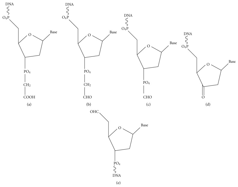
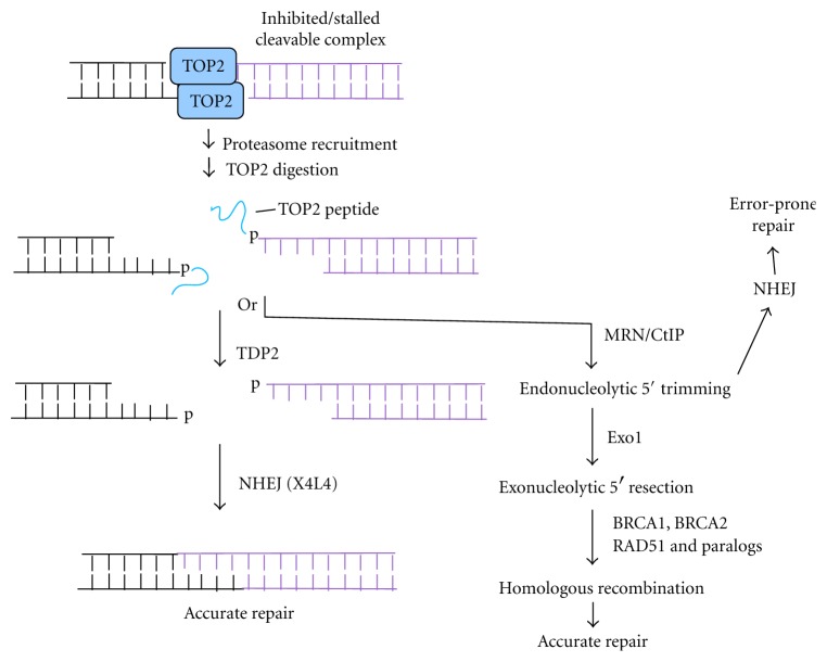
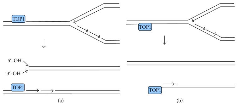
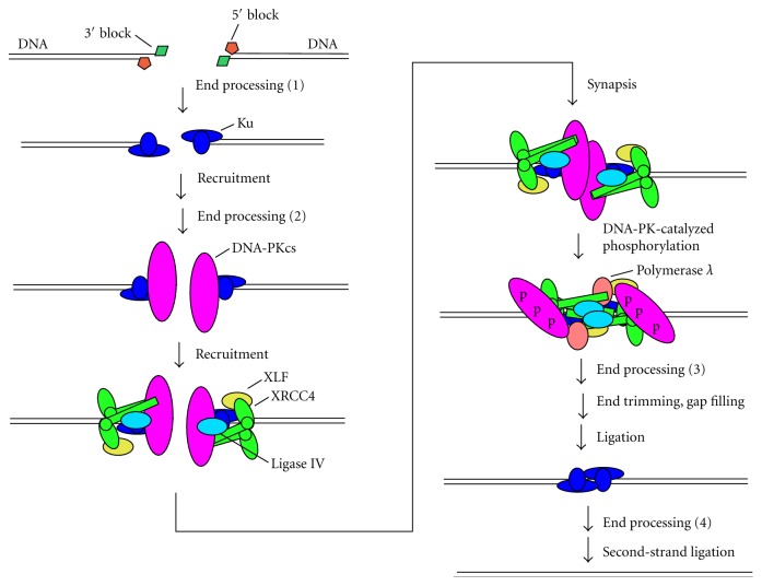
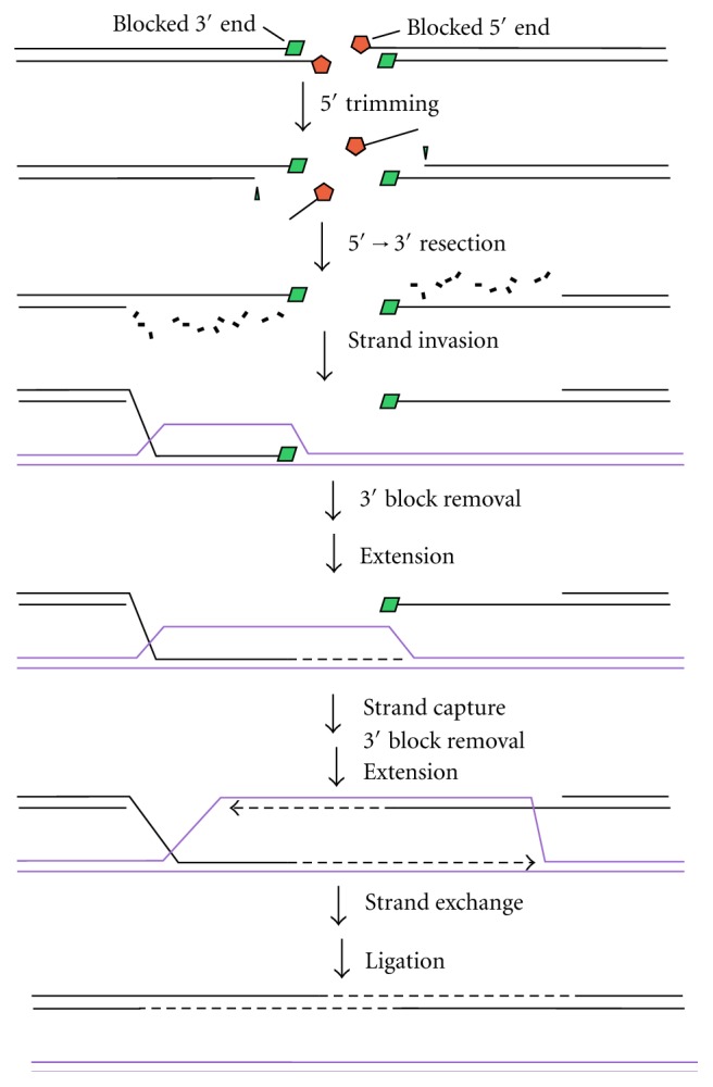
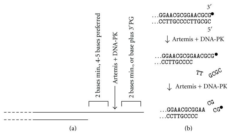
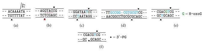
Most DNA double-strand breaks (DSBs)formed in a natural environment have chemical modifications at or near the ends that preclude direct religation and require removal or other processing so that rejoining can proceed. Free radical-mediated DSBs typically bear unligatable 3'-phosphate or 3'-phosphoglycolate termini and often have oxidized bases and/or abasic sites near the break. Topoisomerase-mediated DSBs are blocked by covalently bound peptide fragments of the topoisomerase. Enzymes capable of resolving damaged ends include polynucleotide kinase/phosphatase, which restores missing 5'-phosphates and removes 3'-phosphates; tyrosyl-DNA phosphodiesterases I and II (TDP1 and TDP2), which remove peptide fragments of topoisomerases I and II, respectively, and the Artemis and Metnase endonucleases, which can trim damaged overhangs of diverse structure. TDP1 as well as APE1 can remove 3'-phosphoglycolates and other 3' blocks, while CtIP appears to provide an alternative pathway for topoisomerase II fragment removal. Ku, a core DSB joining protein, can cleave abasic sites near DNA ends. The downstream processes of patching and ligation are tolerant of residual damage, and can sometimes proceed without complete damage removal. Despite these redundant pathways for resolution, damaged ends appear to be a significant barrier to rejoining, and their resolution may be a rate-limiting step in repair of some DSBs..


Figures

Figure 1
Figure 1
Some of the major damaged termini of free radical-mediated DNA strand breaks. (a) 3′-phosphoglycolate (PG). (b) 3′-phosphoglycoaldehyde. (c) 3′-formyl phosphate. (d) 3′-keto-2′-deoxynucleotide. (e) 5′-aldehyde.
Figure 2
Figure 2
Formation and resolution of TOP2-mediated DSB ends. Proteasome recruitment to a persistent TOP2 cleavable complex results in digestion of covalently linked TOP2 to a short peptide, which can be removed by TDP2 to leave cohesive ligatable ends that can be accurately rejoined by NHEJ. Alternatively, CtIP-dependent endonucleolytic cleavage, perhaps catalyzed by MRE11 in its complex with RAD50 and NBS1 (MRN), can lead to either error-prone NHEJ of the resulting noncohesive ends or to 5′→3′ resection by exonuclease 1, culminating in HRR.
Figure 3
Figure 3
Formation of one-ended DSBs by collision of replication forks with persistent TOP1 cleavable complexes. (a) Collision with a lesion in the leading template strand results in a DSB with both 5′- and 3′-hydroxyl termini, plus a residual 3′-TOP1-terminated SSB. (b) Collision with a lesion in the lagging template strand results in a one-ended DSB with a TOP1-linked 3′-terminus and an Okazaki fragment at the 5′-terminus.
Figure 4
Figure 4
Resolution of damaged termini in NHEJ. Ku binds to DNA ends and recruits DNA-PKcs, the XRCC4/DNA ligase IV complex, and XLF. Synapsis of two DSB ends triggers DNA-PKcs autophosphorylation in trans, inducing a conformational change that allows gap filling by polymerase λ and finally ligation by DNA ligase IV. Removal of 5′- and 3′-terminal blocks by end processing enzymes can potentially occur at several steps in the pathway: before recruitment of any NHEJ factors (1); after recruitment of Ku (2) (e.g., by Ku itself [18]); or after DNA-PKcs autophosphorylation (3), which increases accessibility of the ends. In addition, processing of terminal blocks in the second strand can occur after ligation of the first strand (4), possibly by SSB repair pathways after dissociation of NHEJ proteins.
Figure 5
Figure 5
Resolution of damaged termini for HRR. In HRR, following its initiation by MRN/CtIP, 5′→3′ exonucleolytic resection generates 3′-overhangs, one of which invades a sister duplex of the same sequence. Extension of this 3′-end (dashed line) enlarges the D-loop until it can be captured by the other 3′-overhang. Extension of this overhang followed by resolution of crossover structures restores an intact DNA duplex. Blocked 5′-termini must be removed before 5′→3′ exonucleolytic resection, probably by CtIP-dependent endonuclease activity. Removal of blocked 3′-termini could occur either before or after strand exchange but must occur before the respective extension steps.
Figure 6
Figure 6
Trimming of DNA ends by Artemis nuclease. (a) Substrate specificity for trimming of 3′-overhangs. Long overhangs are trimmed 4-5 bases from the single-strand/double-strand junction. Shorter overhangs can be trimmed to as little as 2 bases, but 2 unpaired bases are required on each side of the cleavage site. A 3′-PG can substitute for the 3′-terminal base. (b) Trimming of blunt ends by Artemis. Several bases are first removed endonucleolytically from the 5′-terminal strand (which can be either phosphate- or hydroxyl terminated), generating a 3′-overhang that is then also trimmed, whether it is terminated in a 3′-PG (∙) or a 3′-hydroxyl. All endonucleolytic trimming by Artemis requires autophosphorylated DNA-PK.
Figure 7
Figure 7
Tolerance for mismatched and damaged ends in patching and ligation for NHEJ. Black text shows sequences of ends prior to joining, while blue text shows fill-in of bases by DNA polymerase λ. Lines above and below the text represent the DNA backbone, triangles show sites of ligation by X4L4, and red bars between the bases show mismatches. All sequences read 5′→3′ in the top strand and 3′→5′ in the bottom strand. (a) Cohesive overhangs with an unpaired 3′-flap, ligated by X4L4 alone [19]. (b) Completely mismatched overhangs ligated in one strand only, by purified X4L4 plus Ku, DNA-PKcs, and XLF in vitro [20]. (c) Patching and ligation of annealed overhangs of two I-SceI-induced DSBs in hamster cells, despite an internal A-A mismatch, inferred from sequencing of repair joints [21]. (d) Ligation of a mismatched duplex resulting from error-prone fill-in in HeLa cell extracts supplemented with X4L4 and a highly error-prone mutant of polymerase λ [22]. The single dash in the top strand sequence indicates a single-base deletion. (e) Patching and ligation of partially complementary overhangs, one of which contains a 3′-terminal 8-oxoG, in X4L4-supplemented HeLa nuclear extracts [23]. (f) Patching and ligation of partially complementary overhangs in one strand, despite persistence of a 3′-PG-terminated break in the opposite strand, also in X4L4-supplemented HeLa nuclear extracts [24].

Similar articles

Cited by

References

    1. Hutchinson F. Chemical changes induced in DNA by ionizing radiation. Progress in Nucleic Acid Research and Molecular Biology. 1985;32:115–154. doi: 10.1016/S0079-6603(08)60347-5. - DOI - PubMed
    1. Dedon P. C., Goldberg I. H. Free-radical mechanisms involved in the formation of sequence-dependent bistranded DNA lesions by the antitumor antibiotics bleomycin, neocarzinostatin, and calicheamicin. Chemical Research in Toxicology. 1992;5(3):311–332. - PubMed
    1. Povirk L. F. DNA damage and mutagenesis by radiomimetic DNA-cleaving agents: bleomycin, neocarzinostatin and other enediynes. Mutation Research. 1996;355(1-2):71–89. doi: 10.1016/0027-5107(96)00023-1. - DOI - PubMed
    1. Cadet J., Ravanat J. L., Tavernaporro M., Menoni H., Angelov D. Oxidatively generated complex DNA damage: tandem and clustered lesions. Cancer Letters. 2012;327(1-2):5–15. doi: 10.1016/j.canlet.2012.04.005. - DOI - PubMed
    1. Iliakis G. E., Pantelias G. E., Okayasu R., Blakely W. F. Induction by H2O2 of DNA and interphase chromosome damage in plateau- phase Chinese hamster ovary cells. Radiation Research. 1992;131(2):192–203. - PubMed

LinkOut - more resources